Calcium »
PDB 4cue-4dfs »
4cuo »
Calcium in PDB 4cuo: Banyan Peroxidase with Glycosylation
Enzymatic activity of Banyan Peroxidase with Glycosylation
All present enzymatic activity of Banyan Peroxidase with Glycosylation:
1.11.1.7;
1.11.1.7;
Protein crystallography data
The structure of Banyan Peroxidase with Glycosylation, PDB code: 4cuo
was solved by
G.J.Palm,
A.Sharma,
W.Hinrichs,
with X-Ray Crystallography technique. A brief refinement statistics is given in the table below:
| Resolution Low / High (Å) | 63.32 / 1.67 |
| Space group | P 32 2 1 |
| Cell size a, b, c (Å), α, β, γ (°) | 73.115, 73.115, 164.596, 90.00, 90.00, 120.00 |
| R / Rfree (%) | 15.654 / 18.029 |
Other elements in 4cuo:
The structure of Banyan Peroxidase with Glycosylation also contains other interesting chemical elements:
| Iron | (Fe) | 1 atom |
| Chlorine | (Cl) | 3 atoms |
| Sodium | (Na) | 1 atom |
Calcium Binding Sites:
The binding sites of Calcium atom in the Banyan Peroxidase with Glycosylation
(pdb code 4cuo). This binding sites where shown within
5.0 Angstroms radius around Calcium atom.
In total 2 binding sites of Calcium where determined in the Banyan Peroxidase with Glycosylation, PDB code: 4cuo:
Jump to Calcium binding site number: 1; 2;
In total 2 binding sites of Calcium where determined in the Banyan Peroxidase with Glycosylation, PDB code: 4cuo:
Jump to Calcium binding site number: 1; 2;
Calcium binding site 1 out of 2 in 4cuo
Go back to
Calcium binding site 1 out
of 2 in the Banyan Peroxidase with Glycosylation
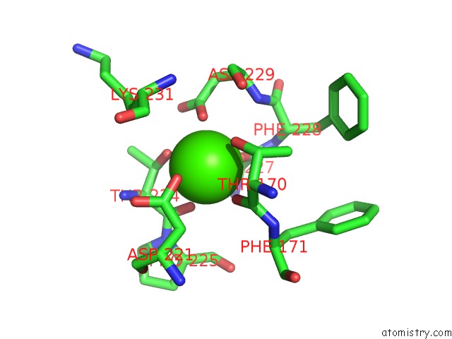
Mono view
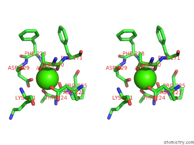
Stereo pair view
Mono view
Stereo pair view
A full contact list of Calcium with other atoms in the Ca binding
site number 1 of Banyan Peroxidase with Glycosylation within 5.0Å range:
|
Calcium binding site 2 out of 2 in 4cuo
Go back to
Calcium binding site 2 out
of 2 in the Banyan Peroxidase with Glycosylation
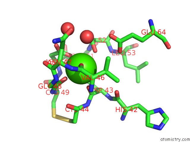
Mono view
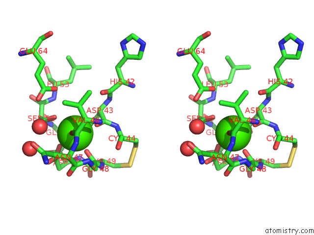
Stereo pair view
Mono view
Stereo pair view
A full contact list of Calcium with other atoms in the Ca binding
site number 2 of Banyan Peroxidase with Glycosylation within 5.0Å range:
|
Reference:
G.J.Palm,
A.Sharma,
M.Kumari,
S.Panjikar,
D.Albrecht,
M.V.Jagannadham,
W.Hinrichs.
Post-Translational Modification and Extended Glycosylation Pattern of A Plant Latex Peroxidase of Native Source Characterized By X-Ray Crystallography. Febs J. V. 281 4319 2014.
ISSN: ISSN 1742-464X
PubMed: 24980207
DOI: 10.1111/FEBS.12900
Page generated: Sat Jul 13 23:17:47 2024
ISSN: ISSN 1742-464X
PubMed: 24980207
DOI: 10.1111/FEBS.12900
Last articles
Zn in 9MJ5Zn in 9HNW
Zn in 9G0L
Zn in 9FNE
Zn in 9DZN
Zn in 9E0I
Zn in 9D32
Zn in 9DAK
Zn in 8ZXC
Zn in 8ZUF