Calcium »
PDB 4dg4-4dtj »
4djc »
Calcium in PDB 4djc: 1.35 A Crystal Structure of the NAV1.5 Diii-IV-Ca/Cam Complex
Protein crystallography data
The structure of 1.35 A Crystal Structure of the NAV1.5 Diii-IV-Ca/Cam Complex, PDB code: 4djc
was solved by
M.F.Sarhan,
C.-C.Tung,
F.Van Petegem,
C.A.Ahern,
with X-Ray Crystallography technique. A brief refinement statistics is given in the table below:
| Resolution Low / High (Å) | 29.55 / 1.35 |
| Space group | P 21 21 21 |
| Cell size a, b, c (Å), α, β, γ (°) | 28.380, 72.720, 97.010, 90.00, 90.00, 90.00 |
| R / Rfree (%) | 15.3 / 17.6 |
Calcium Binding Sites:
The binding sites of Calcium atom in the 1.35 A Crystal Structure of the NAV1.5 Diii-IV-Ca/Cam Complex
(pdb code 4djc). This binding sites where shown within
5.0 Angstroms radius around Calcium atom.
In total 4 binding sites of Calcium where determined in the 1.35 A Crystal Structure of the NAV1.5 Diii-IV-Ca/Cam Complex, PDB code: 4djc:
Jump to Calcium binding site number: 1; 2; 3; 4;
In total 4 binding sites of Calcium where determined in the 1.35 A Crystal Structure of the NAV1.5 Diii-IV-Ca/Cam Complex, PDB code: 4djc:
Jump to Calcium binding site number: 1; 2; 3; 4;
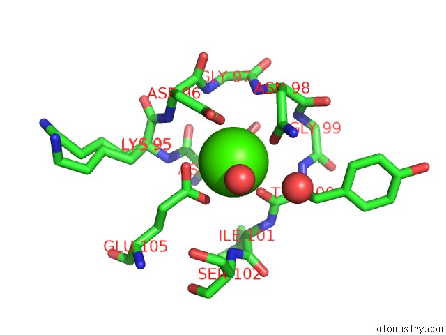
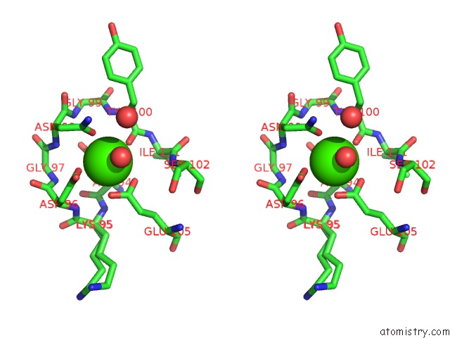
Calcium binding site 1 out of 4 in 4djc
Go back to
Calcium binding site 1 out
of 4 in the 1.35 A Crystal Structure of the NAV1.5 Diii-IV-Ca/Cam Complex
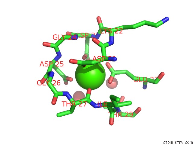
Mono view
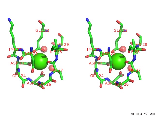
Stereo pair view
Mono view
Stereo pair view
A full contact list of Calcium with other atoms in the Ca binding
site number 1 of 1.35 A Crystal Structure of the NAV1.5 Diii-IV-Ca/Cam Complex within 5.0Å range:
|
Calcium binding site 2 out of 4 in 4djc
Go back to
Calcium binding site 2 out
of 4 in the 1.35 A Crystal Structure of the NAV1.5 Diii-IV-Ca/Cam Complex
Mono view
Stereo pair view
Mono view
Stereo pair view
A full contact list of Calcium with other atoms in the Ca binding
site number 2 of 1.35 A Crystal Structure of the NAV1.5 Diii-IV-Ca/Cam Complex within 5.0Å range:
|
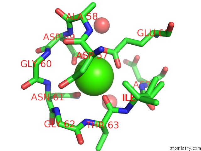
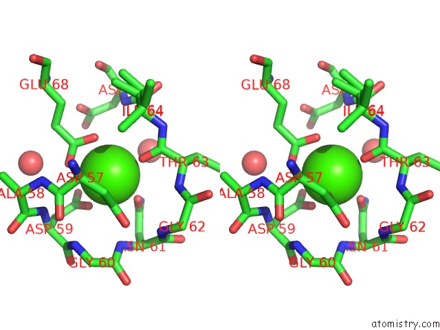
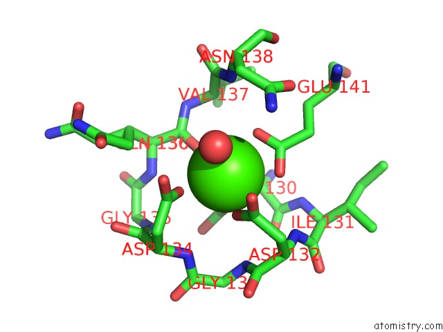
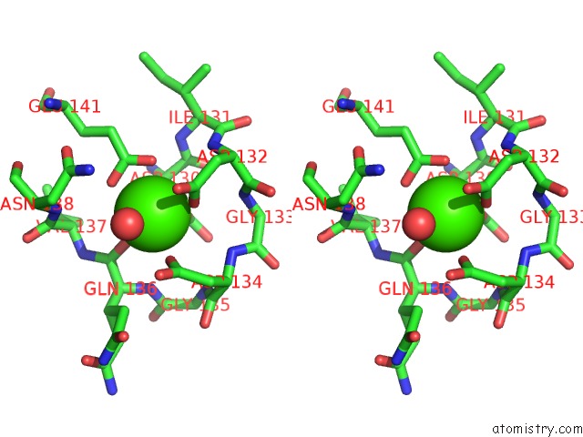
Calcium binding site 3 out of 4 in 4djc
Go back to
Calcium binding site 3 out
of 4 in the 1.35 A Crystal Structure of the NAV1.5 Diii-IV-Ca/Cam Complex
Mono view
Stereo pair view
Mono view
Stereo pair view
A full contact list of Calcium with other atoms in the Ca binding
site number 3 of 1.35 A Crystal Structure of the NAV1.5 Diii-IV-Ca/Cam Complex within 5.0Å range:
|
Calcium binding site 4 out of 4 in 4djc
Go back to
Calcium binding site 4 out
of 4 in the 1.35 A Crystal Structure of the NAV1.5 Diii-IV-Ca/Cam Complex
Mono view
Stereo pair view
Mono view
Stereo pair view
A full contact list of Calcium with other atoms in the Ca binding
site number 4 of 1.35 A Crystal Structure of the NAV1.5 Diii-IV-Ca/Cam Complex within 5.0Å range:
|
Reference:
M.F.Sarhan,
C.C.Tung,
F.Van Petegem,
C.A.Ahern.
Crystallographic Basis For Calcium Regulation of Sodium Channels. Proc.Natl.Acad.Sci.Usa V. 109 3558 2012.
ISSN: ISSN 0027-8424
PubMed: 22331908
DOI: 10.1073/PNAS.1114748109
Page generated: Tue Jul 8 19:26:58 2025
ISSN: ISSN 0027-8424
PubMed: 22331908
DOI: 10.1073/PNAS.1114748109
Last articles
F in 4CEXF in 4CDT
F in 4CC6
F in 4CD0
F in 4CCU
F in 4CCB
F in 4CBT
F in 4CAV
F in 4CAP
F in 4CAR