Calcium »
PDB 4dg4-4dtj »
4dk4 »
Calcium in PDB 4dk4: Crystal Structure of Trypanosoma Brucei Dutpase with Dupnp, CA2+ and Na+
Enzymatic activity of Crystal Structure of Trypanosoma Brucei Dutpase with Dupnp, CA2+ and Na+
All present enzymatic activity of Crystal Structure of Trypanosoma Brucei Dutpase with Dupnp, CA2+ and Na+:
3.6.1.23;
3.6.1.23;
Protein crystallography data
The structure of Crystal Structure of Trypanosoma Brucei Dutpase with Dupnp, CA2+ and Na+, PDB code: 4dk4
was solved by
G.R.Hemsworth,
D.Gonzalez-Pacanowska,
K.S.Wilson,
with X-Ray Crystallography technique. A brief refinement statistics is given in the table below:
| Resolution Low / High (Å) | 61.45 / 1.90 |
| Space group | P 21 21 21 |
| Cell size a, b, c (Å), α, β, γ (°) | 58.840, 83.190, 91.180, 90.00, 90.00, 90.00 |
| R / Rfree (%) | 17.7 / 23.2 |
Other elements in 4dk4:
The structure of Crystal Structure of Trypanosoma Brucei Dutpase with Dupnp, CA2+ and Na+ also contains other interesting chemical elements:
| Sodium | (Na) | 2 atoms |
Calcium Binding Sites:
The binding sites of Calcium atom in the Crystal Structure of Trypanosoma Brucei Dutpase with Dupnp, CA2+ and Na+
(pdb code 4dk4). This binding sites where shown within
5.0 Angstroms radius around Calcium atom.
In total 4 binding sites of Calcium where determined in the Crystal Structure of Trypanosoma Brucei Dutpase with Dupnp, CA2+ and Na+, PDB code: 4dk4:
Jump to Calcium binding site number: 1; 2; 3; 4;
In total 4 binding sites of Calcium where determined in the Crystal Structure of Trypanosoma Brucei Dutpase with Dupnp, CA2+ and Na+, PDB code: 4dk4:
Jump to Calcium binding site number: 1; 2; 3; 4;
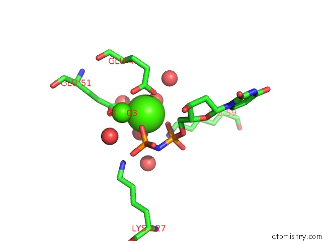
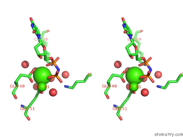
Calcium binding site 1 out of 4 in 4dk4
Go back to
Calcium binding site 1 out
of 4 in the Crystal Structure of Trypanosoma Brucei Dutpase with Dupnp, CA2+ and Na+
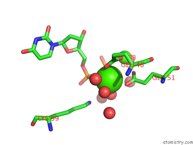
Mono view
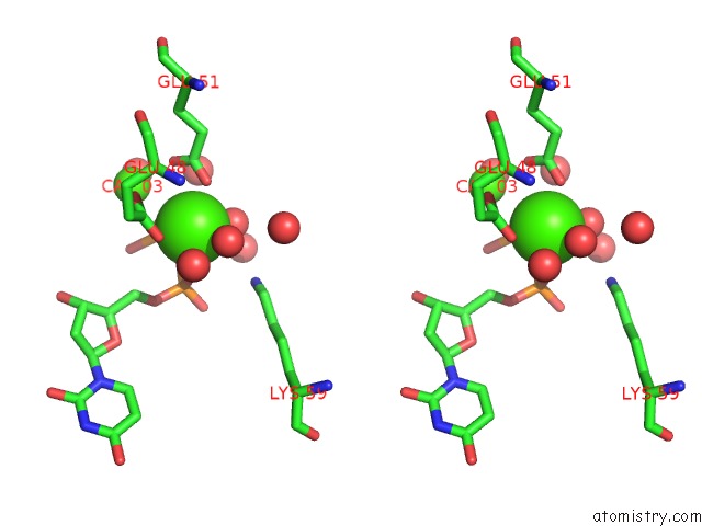
Stereo pair view
Mono view
Stereo pair view
A full contact list of Calcium with other atoms in the Ca binding
site number 1 of Crystal Structure of Trypanosoma Brucei Dutpase with Dupnp, CA2+ and Na+ within 5.0Å range:
|
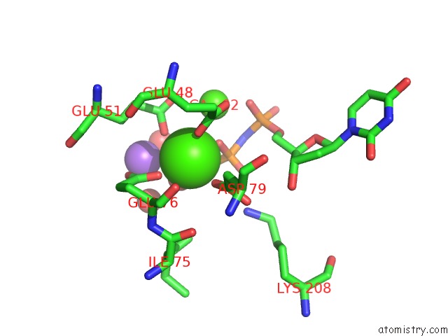
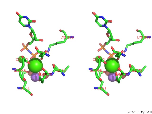
Calcium binding site 2 out of 4 in 4dk4
Go back to
Calcium binding site 2 out
of 4 in the Crystal Structure of Trypanosoma Brucei Dutpase with Dupnp, CA2+ and Na+
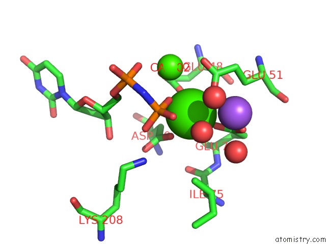
Mono view
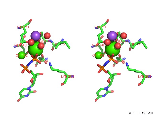
Stereo pair view
Mono view
Stereo pair view
A full contact list of Calcium with other atoms in the Ca binding
site number 2 of Crystal Structure of Trypanosoma Brucei Dutpase with Dupnp, CA2+ and Na+ within 5.0Å range:
|
Calcium binding site 3 out of 4 in 4dk4
Go back to
Calcium binding site 3 out
of 4 in the Crystal Structure of Trypanosoma Brucei Dutpase with Dupnp, CA2+ and Na+
Mono view
Stereo pair view
Mono view
Stereo pair view
A full contact list of Calcium with other atoms in the Ca binding
site number 3 of Crystal Structure of Trypanosoma Brucei Dutpase with Dupnp, CA2+ and Na+ within 5.0Å range:
|
Calcium binding site 4 out of 4 in 4dk4
Go back to
Calcium binding site 4 out
of 4 in the Crystal Structure of Trypanosoma Brucei Dutpase with Dupnp, CA2+ and Na+
Mono view
Stereo pair view
Mono view
Stereo pair view
A full contact list of Calcium with other atoms in the Ca binding
site number 4 of Crystal Structure of Trypanosoma Brucei Dutpase with Dupnp, CA2+ and Na+ within 5.0Å range:
|
Reference:
G.R.Hemsworth,
D.Gonzalez-Pacanowska,
K.S.Wilson.
On the Catalytic Mechanism of Dimeric Dutpases. Biochem.J. V. 456 81 2013.
ISSN: ISSN 0264-6021
PubMed: 24001052
DOI: 10.1042/BJ20130796
Page generated: Sat Jul 13 23:28:29 2024
ISSN: ISSN 0264-6021
PubMed: 24001052
DOI: 10.1042/BJ20130796
Last articles
Zn in 9J0NZn in 9J0O
Zn in 9J0P
Zn in 9FJX
Zn in 9EKB
Zn in 9C0F
Zn in 9CAH
Zn in 9CH0
Zn in 9CH3
Zn in 9CH1