Calcium »
PDB 4dg4-4dtj »
4doq »
Calcium in PDB 4doq: Crystal Structure of the Complex of Porcine Pancreatic Trypsin with 1/2SLPI
Enzymatic activity of Crystal Structure of the Complex of Porcine Pancreatic Trypsin with 1/2SLPI
All present enzymatic activity of Crystal Structure of the Complex of Porcine Pancreatic Trypsin with 1/2SLPI:
3.4.21.4;
3.4.21.4;
Protein crystallography data
The structure of Crystal Structure of the Complex of Porcine Pancreatic Trypsin with 1/2SLPI, PDB code: 4doq
was solved by
K.Fukushima,
M.Takimoto-Kamimura,
with X-Ray Crystallography technique. A brief refinement statistics is given in the table below:
| Resolution Low / High (Å) | 50.06 / 2.00 |
| Space group | P 1 21 1 |
| Cell size a, b, c (Å), α, β, γ (°) | 40.535, 118.610, 93.393, 90.00, 90.74, 90.00 |
| R / Rfree (%) | 18.7 / 24.2 |
Calcium Binding Sites:
The binding sites of Calcium atom in the Crystal Structure of the Complex of Porcine Pancreatic Trypsin with 1/2SLPI
(pdb code 4doq). This binding sites where shown within
5.0 Angstroms radius around Calcium atom.
In total 3 binding sites of Calcium where determined in the Crystal Structure of the Complex of Porcine Pancreatic Trypsin with 1/2SLPI, PDB code: 4doq:
Jump to Calcium binding site number: 1; 2; 3;
In total 3 binding sites of Calcium where determined in the Crystal Structure of the Complex of Porcine Pancreatic Trypsin with 1/2SLPI, PDB code: 4doq:
Jump to Calcium binding site number: 1; 2; 3;
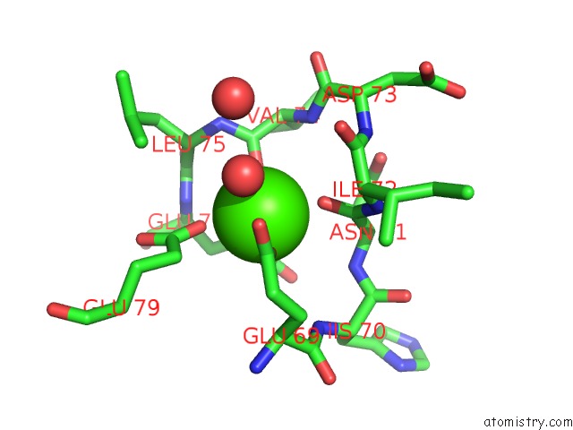
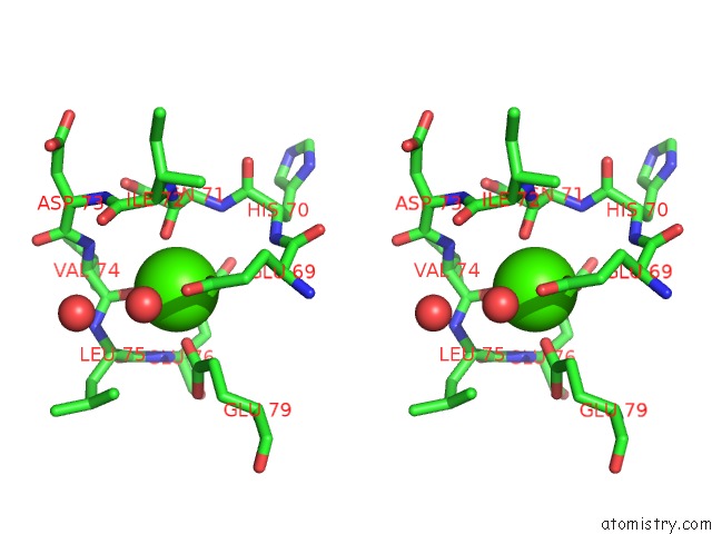
Calcium binding site 1 out of 3 in 4doq
Go back to
Calcium binding site 1 out
of 3 in the Crystal Structure of the Complex of Porcine Pancreatic Trypsin with 1/2SLPI
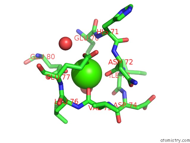
Mono view
Stereo pair view
Mono view
Stereo pair view
A full contact list of Calcium with other atoms in the Ca binding
site number 1 of Crystal Structure of the Complex of Porcine Pancreatic Trypsin with 1/2SLPI within 5.0Å range:
|
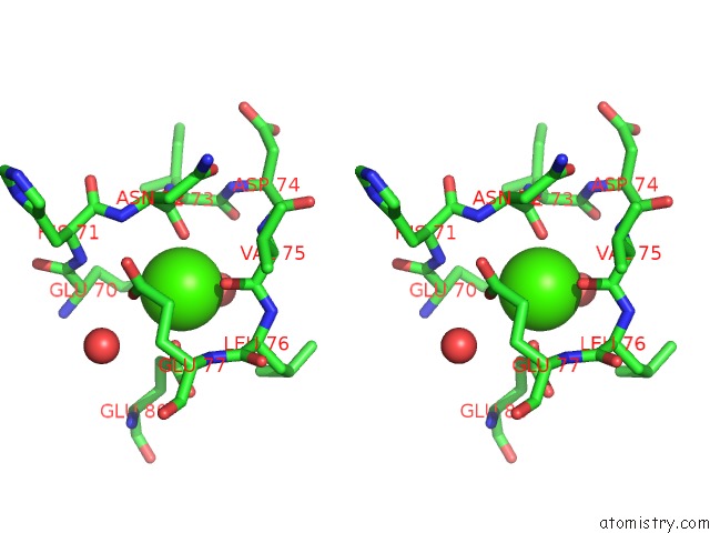
Calcium binding site 2 out of 3 in 4doq
Go back to
Calcium binding site 2 out
of 3 in the Crystal Structure of the Complex of Porcine Pancreatic Trypsin with 1/2SLPI
Mono view
Stereo pair view
Mono view
Stereo pair view
A full contact list of Calcium with other atoms in the Ca binding
site number 2 of Crystal Structure of the Complex of Porcine Pancreatic Trypsin with 1/2SLPI within 5.0Å range:
|
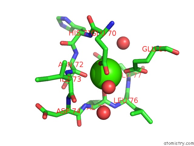
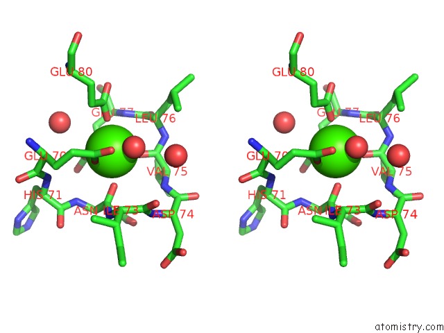
Calcium binding site 3 out of 3 in 4doq
Go back to
Calcium binding site 3 out
of 3 in the Crystal Structure of the Complex of Porcine Pancreatic Trypsin with 1/2SLPI
Mono view
Stereo pair view
Mono view
Stereo pair view
A full contact list of Calcium with other atoms in the Ca binding
site number 3 of Crystal Structure of the Complex of Porcine Pancreatic Trypsin with 1/2SLPI within 5.0Å range:
|
Reference:
K.Fukushima,
T.Kamimura,
M.Takimoto-Kamimura.
Structure Basis 1/2SLPI and Porcine Pancreas Trypsin Interaction J.Synchrotron Radiat. V. 20 943 2013.
ISSN: ISSN 0909-0495
PubMed: 24121345
DOI: 10.1107/S090904951302133X
Page generated: Sat Jul 13 23:34:39 2024
ISSN: ISSN 0909-0495
PubMed: 24121345
DOI: 10.1107/S090904951302133X
Last articles
Zn in 9J0NZn in 9J0O
Zn in 9J0P
Zn in 9FJX
Zn in 9EKB
Zn in 9C0F
Zn in 9CAH
Zn in 9CH0
Zn in 9CH3
Zn in 9CH1