Calcium »
PDB 4dg4-4dtj »
4dsj »
Calcium in PDB 4dsj: Crystal Structure of Fragment Dna Polymerase I From Bacillus Stearothermophilus with Duplex Dna, Dgtp and Calcium
Enzymatic activity of Crystal Structure of Fragment Dna Polymerase I From Bacillus Stearothermophilus with Duplex Dna, Dgtp and Calcium
All present enzymatic activity of Crystal Structure of Fragment Dna Polymerase I From Bacillus Stearothermophilus with Duplex Dna, Dgtp and Calcium:
2.7.7.7;
2.7.7.7;
Protein crystallography data
The structure of Crystal Structure of Fragment Dna Polymerase I From Bacillus Stearothermophilus with Duplex Dna, Dgtp and Calcium, PDB code: 4dsj
was solved by
J.H.Gan,
R.Abdur,
H.H.Liu,
J.Sheng,
J.Caton-Willians,
A.S.Soares,
Z.Huang,
with X-Ray Crystallography technique. A brief refinement statistics is given in the table below:
| Resolution Low / High (Å) | 114.73 / 2.86 |
| Space group | P 21 21 21 |
| Cell size a, b, c (Å), α, β, γ (°) | 60.016, 112.070, 229.450, 90.00, 90.00, 90.00 |
| R / Rfree (%) | 21.3 / 27.7 |
Calcium Binding Sites:
The binding sites of Calcium atom in the Crystal Structure of Fragment Dna Polymerase I From Bacillus Stearothermophilus with Duplex Dna, Dgtp and Calcium
(pdb code 4dsj). This binding sites where shown within
5.0 Angstroms radius around Calcium atom.
In total 2 binding sites of Calcium where determined in the Crystal Structure of Fragment Dna Polymerase I From Bacillus Stearothermophilus with Duplex Dna, Dgtp and Calcium, PDB code: 4dsj:
Jump to Calcium binding site number: 1; 2;
In total 2 binding sites of Calcium where determined in the Crystal Structure of Fragment Dna Polymerase I From Bacillus Stearothermophilus with Duplex Dna, Dgtp and Calcium, PDB code: 4dsj:
Jump to Calcium binding site number: 1; 2;
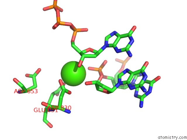
Calcium binding site 1 out of 2 in 4dsj
Go back to
Calcium binding site 1 out
of 2 in the Crystal Structure of Fragment Dna Polymerase I From Bacillus Stearothermophilus with Duplex Dna, Dgtp and Calcium
Mono view

Stereo pair view
Mono view
Stereo pair view
A full contact list of Calcium with other atoms in the Ca binding
site number 1 of Crystal Structure of Fragment Dna Polymerase I From Bacillus Stearothermophilus with Duplex Dna, Dgtp and Calcium within 5.0Å range:
|
Calcium binding site 2 out of 2 in 4dsj
Go back to
Calcium binding site 2 out
of 2 in the Crystal Structure of Fragment Dna Polymerase I From Bacillus Stearothermophilus with Duplex Dna, Dgtp and Calcium

Mono view

Stereo pair view
Mono view
Stereo pair view
A full contact list of Calcium with other atoms in the Ca binding
site number 2 of Crystal Structure of Fragment Dna Polymerase I From Bacillus Stearothermophilus with Duplex Dna, Dgtp and Calcium within 5.0Å range:
|
Reference:
J.H.Gan,
R.Abdur,
H.H.Liu,
J.Sheng,
J.Caton-Willians,
A.S.Soares,
Z.Huang.
Biochemical and Structural Insights Into the Fidelity of Bacillus Stearothermophilus Dna Polymerase To Be Published.
Page generated: Tue Jul 8 19:33:14 2025
Last articles
Cl in 8C9FCl in 8C9G
Cl in 8C9E
Cl in 8C8D
Cl in 8C8S
Cl in 8C8J
Cl in 8C7J
Cl in 8C7Y
Cl in 8C8C
Cl in 8C7X