Calcium »
PDB 4dtm-4ec5 »
4du6 »
Calcium in PDB 4du6: Crystal Structure of Gtp Cyclohydrolase I From Yersinia Pestis Complexed with Gtp
Enzymatic activity of Crystal Structure of Gtp Cyclohydrolase I From Yersinia Pestis Complexed with Gtp
All present enzymatic activity of Crystal Structure of Gtp Cyclohydrolase I From Yersinia Pestis Complexed with Gtp:
3.5.4.16;
3.5.4.16;
Protein crystallography data
The structure of Crystal Structure of Gtp Cyclohydrolase I From Yersinia Pestis Complexed with Gtp, PDB code: 4du6
was solved by
N.Maltseva,
Y.Kim,
K.Kwon,
W.F.Anderson,
A.Joachimiak,
Center Forstructural Genomics Of Infectious Diseases (Csgid),
with X-Ray Crystallography technique. A brief refinement statistics is given in the table below:
| Resolution Low / High (Å) | 43.20 / 2.11 |
| Space group | C 1 2 1 |
| Cell size a, b, c (Å), α, β, γ (°) | 174.047, 104.912, 70.070, 90.00, 96.89, 90.00 |
| R / Rfree (%) | 18 / 22.5 |
Calcium Binding Sites:
The binding sites of Calcium atom in the Crystal Structure of Gtp Cyclohydrolase I From Yersinia Pestis Complexed with Gtp
(pdb code 4du6). This binding sites where shown within
5.0 Angstroms radius around Calcium atom.
In total 3 binding sites of Calcium where determined in the Crystal Structure of Gtp Cyclohydrolase I From Yersinia Pestis Complexed with Gtp, PDB code: 4du6:
Jump to Calcium binding site number: 1; 2; 3;
In total 3 binding sites of Calcium where determined in the Crystal Structure of Gtp Cyclohydrolase I From Yersinia Pestis Complexed with Gtp, PDB code: 4du6:
Jump to Calcium binding site number: 1; 2; 3;
Calcium binding site 1 out of 3 in 4du6
Go back to
Calcium binding site 1 out
of 3 in the Crystal Structure of Gtp Cyclohydrolase I From Yersinia Pestis Complexed with Gtp
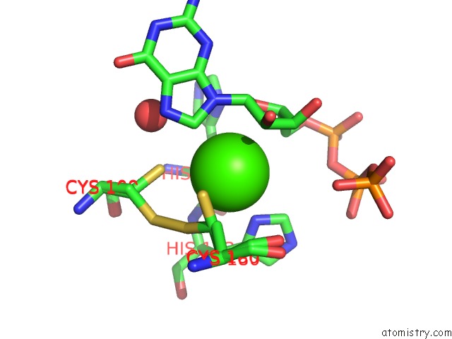
Mono view

Stereo pair view
Mono view
Stereo pair view
A full contact list of Calcium with other atoms in the Ca binding
site number 1 of Crystal Structure of Gtp Cyclohydrolase I From Yersinia Pestis Complexed with Gtp within 5.0Å range:
|
Calcium binding site 2 out of 3 in 4du6
Go back to
Calcium binding site 2 out
of 3 in the Crystal Structure of Gtp Cyclohydrolase I From Yersinia Pestis Complexed with Gtp
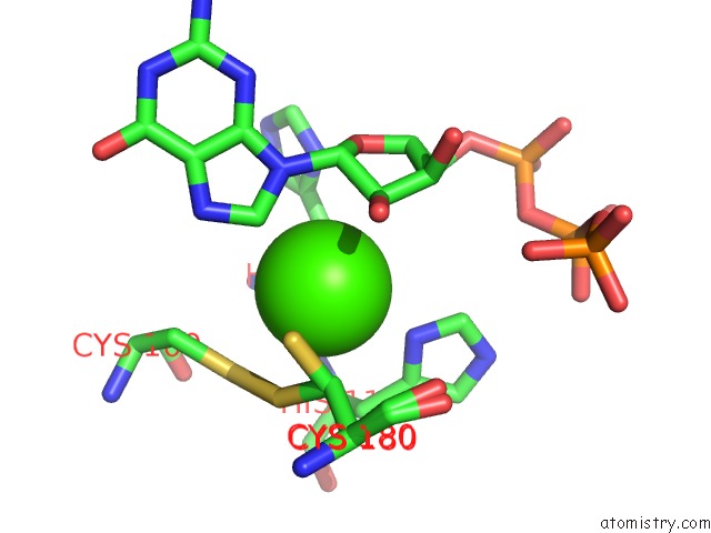
Mono view
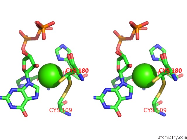
Stereo pair view
Mono view
Stereo pair view
A full contact list of Calcium with other atoms in the Ca binding
site number 2 of Crystal Structure of Gtp Cyclohydrolase I From Yersinia Pestis Complexed with Gtp within 5.0Å range:
|
Calcium binding site 3 out of 3 in 4du6
Go back to
Calcium binding site 3 out
of 3 in the Crystal Structure of Gtp Cyclohydrolase I From Yersinia Pestis Complexed with Gtp

Mono view
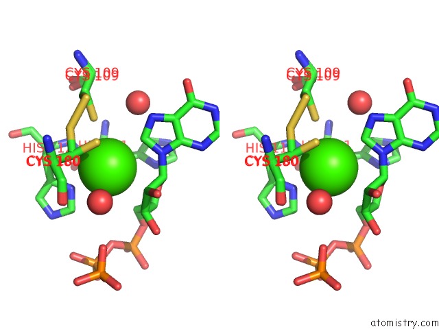
Stereo pair view
Mono view
Stereo pair view
A full contact list of Calcium with other atoms in the Ca binding
site number 3 of Crystal Structure of Gtp Cyclohydrolase I From Yersinia Pestis Complexed with Gtp within 5.0Å range:
|
Reference:
N.Maltseva,
Y.Kim,
K.Kwon,
W.F.Anderson,
A.Joachimiak,
Csgid.
Crystal Structure of Gtp Cyclohydrolase I From Yersinia Pestis Complexed with Gtp To Be Published.
Page generated: Tue Jul 8 19:36:30 2025
Last articles
Cl in 5JSMCl in 5JSJ
Cl in 5JSH
Cl in 5JRD
Cl in 5JSC
Cl in 5JSG
Cl in 5JRG
Cl in 5JSF
Cl in 5JS6
Cl in 5JQG