Calcium »
PDB 4dtm-4ec5 »
4e5u »
Calcium in PDB 4e5u: The Crystal Structure of Thymidylate Kinase From Pseudomonas Aeruginosa PAO1 in Complex with Thymidine Monophosphate.
Enzymatic activity of The Crystal Structure of Thymidylate Kinase From Pseudomonas Aeruginosa PAO1 in Complex with Thymidine Monophosphate.
All present enzymatic activity of The Crystal Structure of Thymidylate Kinase From Pseudomonas Aeruginosa PAO1 in Complex with Thymidine Monophosphate.:
2.7.4.9;
2.7.4.9;
Protein crystallography data
The structure of The Crystal Structure of Thymidylate Kinase From Pseudomonas Aeruginosa PAO1 in Complex with Thymidine Monophosphate., PDB code: 4e5u
was solved by
K.Tan,
G.Joachimiak,
R.Jedrzejczak,
J.Sacchettini,
A.Joachimiak,
Midwestcenter For Structural Genomics (Mcsg),
Structures Of Mtb Proteinsconferring Susceptibility To Known Mtb Inhibitors (Mtbi),
with X-Ray Crystallography technique. A brief refinement statistics is given in the table below:
| Resolution Low / High (Å) | 35.96 / 1.81 |
| Space group | P 21 21 21 |
| Cell size a, b, c (Å), α, β, γ (°) | 46.132, 57.399, 149.952, 90.00, 90.00, 90.00 |
| R / Rfree (%) | 18.8 / 23.1 |
Calcium Binding Sites:
The binding sites of Calcium atom in the The Crystal Structure of Thymidylate Kinase From Pseudomonas Aeruginosa PAO1 in Complex with Thymidine Monophosphate.
(pdb code 4e5u). This binding sites where shown within
5.0 Angstroms radius around Calcium atom.
In total 3 binding sites of Calcium where determined in the The Crystal Structure of Thymidylate Kinase From Pseudomonas Aeruginosa PAO1 in Complex with Thymidine Monophosphate., PDB code: 4e5u:
Jump to Calcium binding site number: 1; 2; 3;
In total 3 binding sites of Calcium where determined in the The Crystal Structure of Thymidylate Kinase From Pseudomonas Aeruginosa PAO1 in Complex with Thymidine Monophosphate., PDB code: 4e5u:
Jump to Calcium binding site number: 1; 2; 3;
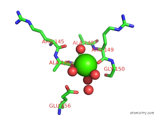
Calcium binding site 1 out of 3 in 4e5u
Go back to
Calcium binding site 1 out
of 3 in the The Crystal Structure of Thymidylate Kinase From Pseudomonas Aeruginosa PAO1 in Complex with Thymidine Monophosphate.
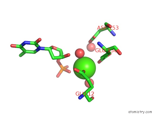
Mono view
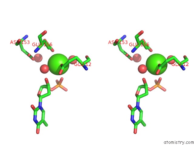
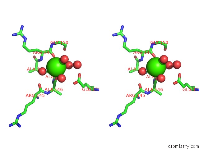
Stereo pair view
Mono view
Stereo pair view
A full contact list of Calcium with other atoms in the Ca binding
site number 1 of The Crystal Structure of Thymidylate Kinase From Pseudomonas Aeruginosa PAO1 in Complex with Thymidine Monophosphate. within 5.0Å range:
|
Calcium binding site 2 out of 3 in 4e5u
Go back to
Calcium binding site 2 out
of 3 in the The Crystal Structure of Thymidylate Kinase From Pseudomonas Aeruginosa PAO1 in Complex with Thymidine Monophosphate.
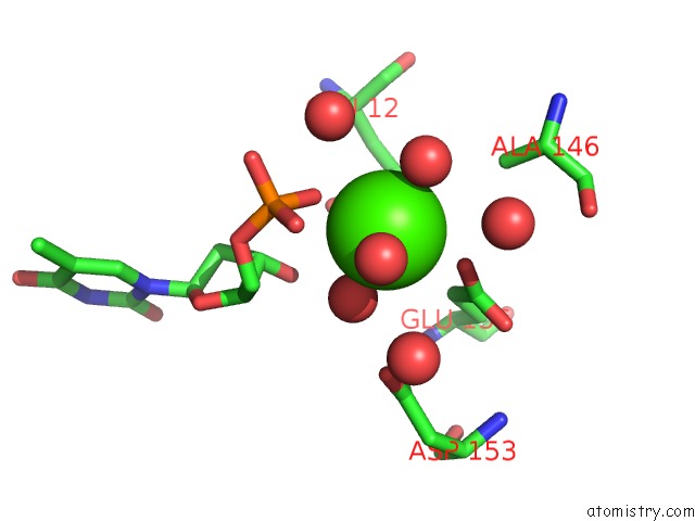
Mono view
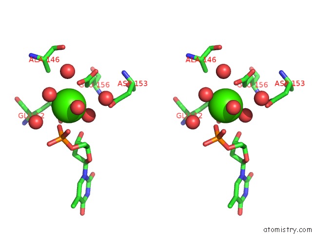
Stereo pair view
Mono view
Stereo pair view
A full contact list of Calcium with other atoms in the Ca binding
site number 2 of The Crystal Structure of Thymidylate Kinase From Pseudomonas Aeruginosa PAO1 in Complex with Thymidine Monophosphate. within 5.0Å range:
|
Calcium binding site 3 out of 3 in 4e5u
Go back to
Calcium binding site 3 out
of 3 in the The Crystal Structure of Thymidylate Kinase From Pseudomonas Aeruginosa PAO1 in Complex with Thymidine Monophosphate.
Mono view
Stereo pair view
Mono view
Stereo pair view
A full contact list of Calcium with other atoms in the Ca binding
site number 3 of The Crystal Structure of Thymidylate Kinase From Pseudomonas Aeruginosa PAO1 in Complex with Thymidine Monophosphate. within 5.0Å range:
|
Reference:
K.Tan,
G.Joachimiak,
R.Jedrzejczak,
J.Sacchettini,
A.Joachimiak.
The Crystal Structure of Thymidylate Kinase From Pseudomonas Aeruginosa PAO1 in Complex with Thymidine Monophosphate. To Be Published.
Page generated: Sat Jul 13 23:46:11 2024
Last articles
Zn in 9J0NZn in 9J0O
Zn in 9J0P
Zn in 9FJX
Zn in 9EKB
Zn in 9C0F
Zn in 9CAH
Zn in 9CH0
Zn in 9CH3
Zn in 9CH1