Calcium »
PDB 4ecg-4enz »
4ecy »
Calcium in PDB 4ecy: Human Dna Polymerase Eta - Dna Ternary Complex: at Crystal at pH 6.0 (Na+ Mes) with 1 CA2+ Ion
Enzymatic activity of Human Dna Polymerase Eta - Dna Ternary Complex: at Crystal at pH 6.0 (Na+ Mes) with 1 CA2+ Ion
All present enzymatic activity of Human Dna Polymerase Eta - Dna Ternary Complex: at Crystal at pH 6.0 (Na+ Mes) with 1 CA2+ Ion:
2.7.7.7;
2.7.7.7;
Protein crystallography data
The structure of Human Dna Polymerase Eta - Dna Ternary Complex: at Crystal at pH 6.0 (Na+ Mes) with 1 CA2+ Ion, PDB code: 4ecy
was solved by
T.Nakamura,
Y.Zhao,
W.Yang,
with X-Ray Crystallography technique. A brief refinement statistics is given in the table below:
| Resolution Low / High (Å) | 28.52 / 1.94 |
| Space group | P 61 |
| Cell size a, b, c (Å), α, β, γ (°) | 98.786, 98.786, 82.431, 90.00, 90.00, 120.00 |
| R / Rfree (%) | 17.9 / 23.2 |
Calcium Binding Sites:
The binding sites of Calcium atom in the Human Dna Polymerase Eta - Dna Ternary Complex: at Crystal at pH 6.0 (Na+ Mes) with 1 CA2+ Ion
(pdb code 4ecy). This binding sites where shown within
5.0 Angstroms radius around Calcium atom.
In total only one binding site of Calcium was determined in the Human Dna Polymerase Eta - Dna Ternary Complex: at Crystal at pH 6.0 (Na+ Mes) with 1 CA2+ Ion, PDB code: 4ecy:
In total only one binding site of Calcium was determined in the Human Dna Polymerase Eta - Dna Ternary Complex: at Crystal at pH 6.0 (Na+ Mes) with 1 CA2+ Ion, PDB code: 4ecy:
Calcium binding site 1 out of 1 in 4ecy
Go back to
Calcium binding site 1 out
of 1 in the Human Dna Polymerase Eta - Dna Ternary Complex: at Crystal at pH 6.0 (Na+ Mes) with 1 CA2+ Ion
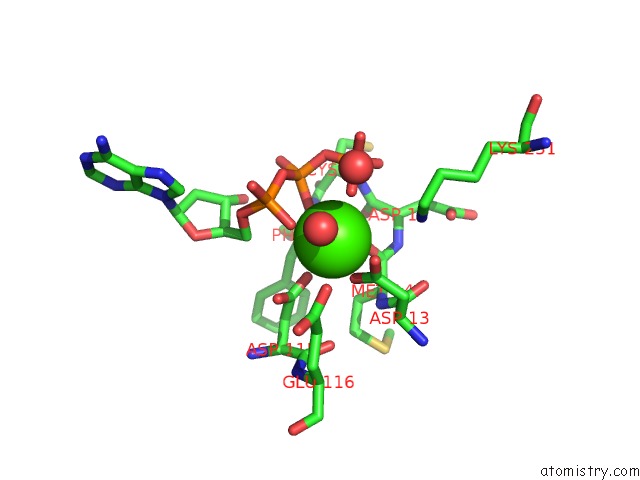
Mono view
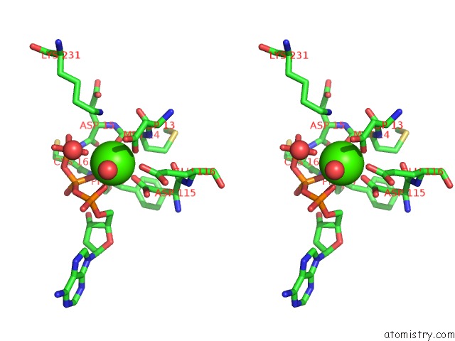
Stereo pair view
Mono view
Stereo pair view
A full contact list of Calcium with other atoms in the Ca binding
site number 1 of Human Dna Polymerase Eta - Dna Ternary Complex: at Crystal at pH 6.0 (Na+ Mes) with 1 CA2+ Ion within 5.0Å range:
|
Reference:
T.Nakamura,
Y.Zhao,
Y.Yamagata,
Y.J.Hua,
W.Yang.
Watching Dna Polymerase Eta Make A Phosphodiester Bond Nature V. 487 196 2012.
ISSN: ISSN 0028-0836
PubMed: 22785315
DOI: 10.1038/NATURE11181
Page generated: Sat Jul 13 23:54:36 2024
ISSN: ISSN 0028-0836
PubMed: 22785315
DOI: 10.1038/NATURE11181
Last articles
Zn in 9J0NZn in 9J0O
Zn in 9J0P
Zn in 9FJX
Zn in 9EKB
Zn in 9C0F
Zn in 9CAH
Zn in 9CH0
Zn in 9CH3
Zn in 9CH1