Calcium »
PDB 4ecg-4enz »
4egd »
Calcium in PDB 4egd: 1.85 Angstrom Crystal Structure of Native Hypothetical Protein SAOUHSC_02783 From Staphylococcus Aureus
Protein crystallography data
The structure of 1.85 Angstrom Crystal Structure of Native Hypothetical Protein SAOUHSC_02783 From Staphylococcus Aureus, PDB code: 4egd
was solved by
M.Biancucci,
G.Minasov,
A.Halavaty,
E.V.Filippova,
L.Shuvalova,
I.Dubrovska,
J.Winsor,
F.Bagnoli,
F.Falugi,
M.Bottomley,
G.Grandi,
W.F.Anderson,
Center For Structural Genomics Of Infectious Diseases(Csgid),
with X-Ray Crystallography technique. A brief refinement statistics is given in the table below:
| Resolution Low / High (Å) | 29.93 / 1.85 |
| Space group | P 21 21 21 |
| Cell size a, b, c (Å), α, β, γ (°) | 44.231, 88.782, 121.622, 90.00, 90.00, 90.00 |
| R / Rfree (%) | 16.8 / 21.5 |
Other elements in 4egd:
The structure of 1.85 Angstrom Crystal Structure of Native Hypothetical Protein SAOUHSC_02783 From Staphylococcus Aureus also contains other interesting chemical elements:
| Chlorine | (Cl) | 1 atom |
Calcium Binding Sites:
The binding sites of Calcium atom in the 1.85 Angstrom Crystal Structure of Native Hypothetical Protein SAOUHSC_02783 From Staphylococcus Aureus
(pdb code 4egd). This binding sites where shown within
5.0 Angstroms radius around Calcium atom.
In total only one binding site of Calcium was determined in the 1.85 Angstrom Crystal Structure of Native Hypothetical Protein SAOUHSC_02783 From Staphylococcus Aureus, PDB code: 4egd:
In total only one binding site of Calcium was determined in the 1.85 Angstrom Crystal Structure of Native Hypothetical Protein SAOUHSC_02783 From Staphylococcus Aureus, PDB code: 4egd:
Calcium binding site 1 out of 1 in 4egd
Go back to
Calcium binding site 1 out
of 1 in the 1.85 Angstrom Crystal Structure of Native Hypothetical Protein SAOUHSC_02783 From Staphylococcus Aureus
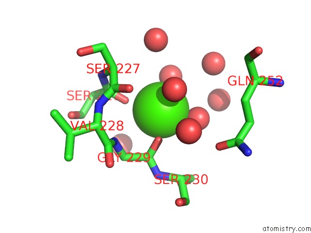
Mono view
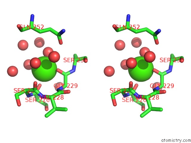
Stereo pair view
Mono view
Stereo pair view
A full contact list of Calcium with other atoms in the Ca binding
site number 1 of 1.85 Angstrom Crystal Structure of Native Hypothetical Protein SAOUHSC_02783 From Staphylococcus Aureus within 5.0Å range:
|
Reference:
M.Biancucci,
G.Minasov,
A.Halavaty,
E.V.Filippova,
L.Shuvalova,
I.Dubrovska,
J.Winsor,
F.Bagnoli,
F.Falugi,
M.Bottomley,
G.Grandi,
W.F.Anderson,
Center For Structural Genomics Of Infectious Diseases(Csgid).
1.85 Angstrom Crystal Structure of Native Hypothetical Protein SAOUHSC_02783 From Staphylococcus Aureus To Be Published.
Page generated: Sat Jul 13 23:56:55 2024
Last articles
Zn in 9JYWZn in 9IR4
Zn in 9IR3
Zn in 9GMX
Zn in 9GMW
Zn in 9JEJ
Zn in 9ERF
Zn in 9ERE
Zn in 9EGV
Zn in 9EGW