Atomistry » Calcium » PDB 4ecg-4enz » 4ej7
Atomistry »
  Calcium »
    PDB 4ecg-4enz »
      4ej7 »

Calcium in PDB 4ej7: Crystal Structure of the Aminoglycoside Phosphotransferase Aph(3')-Ia, Atp-Bound

Enzymatic activity of Crystal Structure of the Aminoglycoside Phosphotransferase Aph(3')-Ia, Atp-Bound

All present enzymatic activity of Crystal Structure of the Aminoglycoside Phosphotransferase Aph(3')-Ia, Atp-Bound:
2.7.1.95;

Protein crystallography data

The structure of Crystal Structure of the Aminoglycoside Phosphotransferase Aph(3')-Ia, Atp-Bound, PDB code: 4ej7 was solved by P.J.Stogios, G.Minasov, K.Tan, E.Evdokimova, O.Egorova, R.Di Leo, T.Shakya, G.D.Wright, A.Savchenko, W.F.Anderson, Center For Structural Genomicsof Infectious Diseases (Csgid), with X-Ray Crystallography technique. A brief refinement statistics is given in the table below:

Resolution Low / High (Å) 43.67 / 2.29
Space group C 2 2 21
Cell size a, b, c (Å), α, β, γ (°) 85.363, 152.466, 165.569, 90.00, 90.00, 90.00
R / Rfree (%) 21.3 / 25.2

Calcium Binding Sites:

Pages:

>>> Page 1 <<< Page 2, Binding sites: 11 - 12;

Binding sites:

The binding sites of Calcium atom in the Crystal Structure of the Aminoglycoside Phosphotransferase Aph(3')-Ia, Atp-Bound (pdb code 4ej7). This binding sites where shown within 5.0 Angstroms radius around Calcium atom.
In total 12 binding sites of Calcium where determined in the Crystal Structure of the Aminoglycoside Phosphotransferase Aph(3')-Ia, Atp-Bound, PDB code: 4ej7:
Jump to Calcium binding site number: 1; 2; 3; 4; 5; 6; 7; 8; 9; 10;

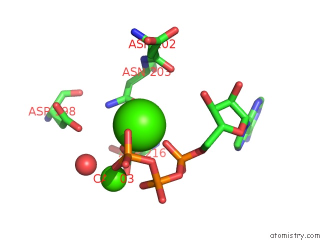
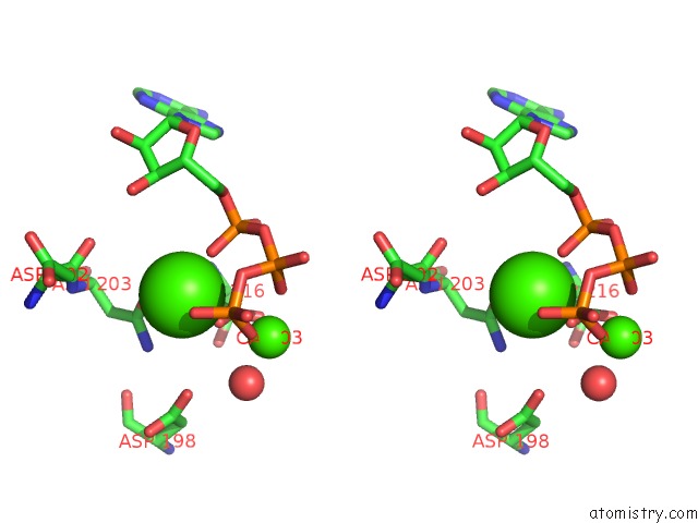
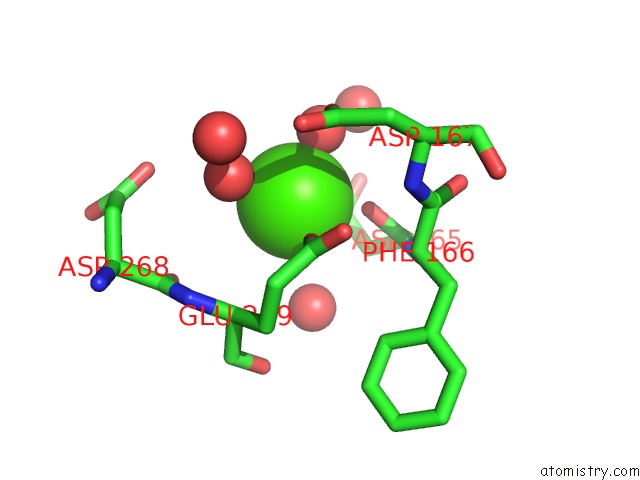
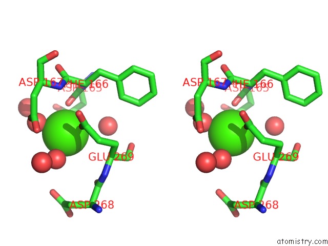
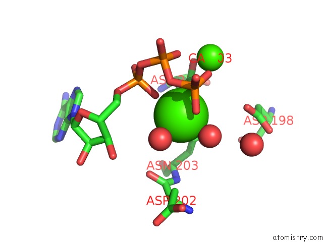
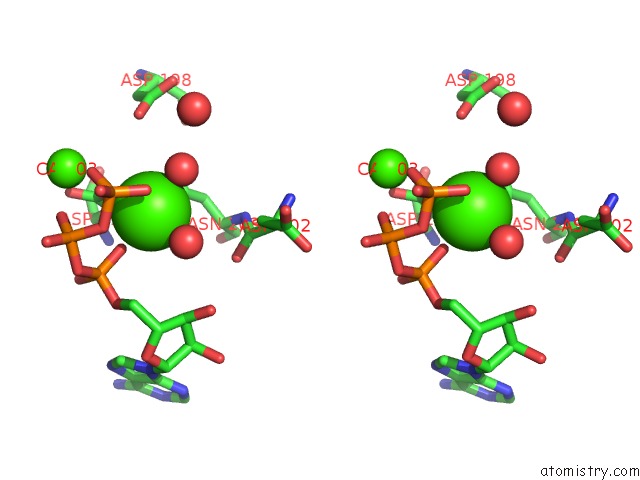
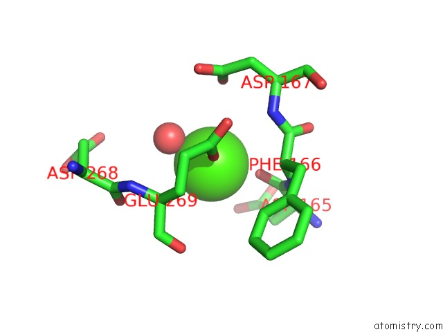
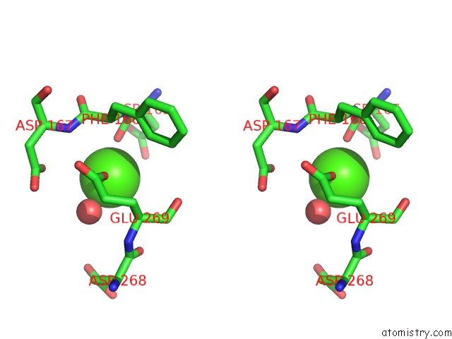
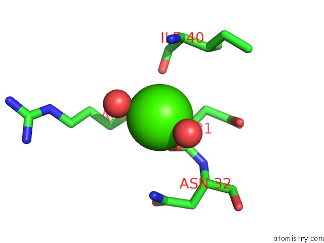
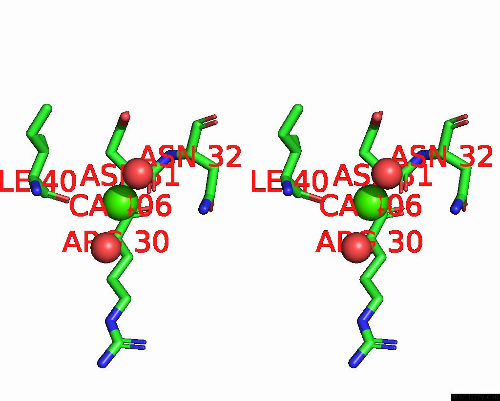
Calcium binding site 1 out of 12 in 4ej7

Go back to Calcium Binding Sites List in 4ej7
Calcium binding site 1 out of 12 in the Crystal Structure of the Aminoglycoside Phosphotransferase Aph(3')-Ia, Atp-Bound


Mono view


Stereo pair view

A full contact list of Calcium with other atoms in the Ca binding site number 1 of Crystal Structure of the Aminoglycoside Phosphotransferase Aph(3')-Ia, Atp-Bound within 5.0Å range:
probe atom residue distance (Å) B Occ
A:Ca402

b:27.9
occ:1.00
O1A A:ATP401 2.2 27.4 1.0
OD1 A:ASN203 2.4 23.3 1.0
O1G A:ATP401 2.5 68.8 1.0
OD2 A:ASP216 2.5 20.4 1.0
PG A:ATP401 3.2 65.0 1.0
O3B A:ATP401 3.2 48.0 1.0
CG A:ASN203 3.4 23.6 1.0
CG A:ASP216 3.4 20.3 1.0
O2G A:ATP401 3.5 59.4 1.0
ND2 A:ASN203 3.6 22.8 1.0
PA A:ATP401 3.7 31.1 1.0
CB A:ASP216 3.8 15.9 1.0
CA A:CA403 4.1 33.2 1.0
O2B A:ATP401 4.1 24.5 1.0
PB A:ATP401 4.2 27.2 1.0
O3A A:ATP401 4.3 43.9 1.0
OD1 A:ASP198 4.4 26.5 1.0
OD1 A:ASP216 4.5 23.1 1.0
O A:ASP202 4.6 32.2 1.0
O5' A:ATP401 4.6 26.2 1.0
O3G A:ATP401 4.7 66.6 1.0
O2A A:ATP401 4.7 33.9 1.0
CB A:ASN203 4.7 25.2 1.0
C5' A:ATP401 4.8 22.9 1.0
C A:ASP202 4.8 26.7 1.0
O3' A:ATP401 4.9 32.5 1.0
O A:HOH599 4.9 29.2 1.0
CB A:ASP202 4.9 41.7 1.0

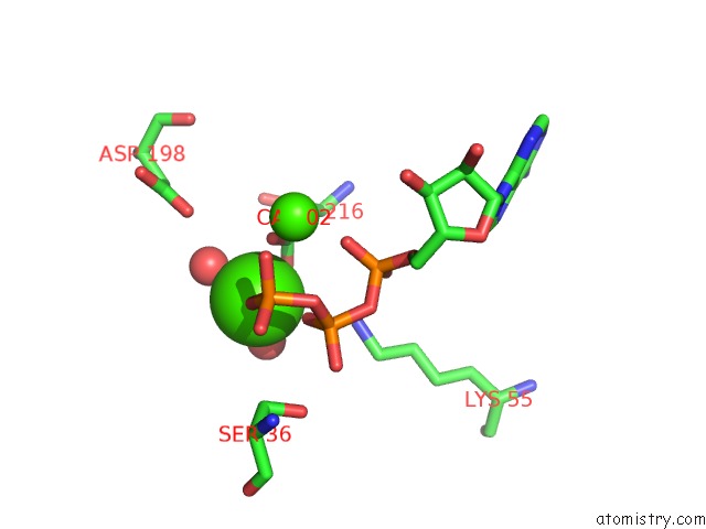
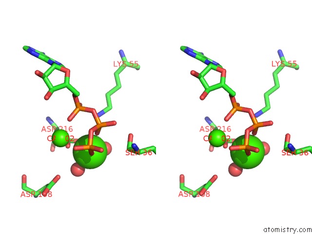
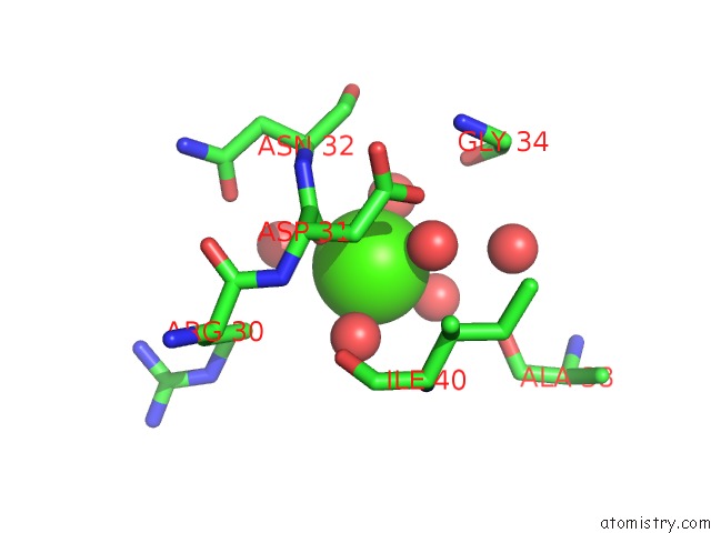
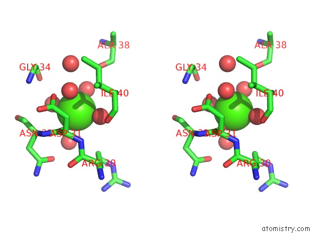
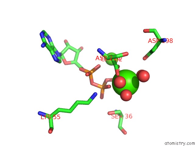
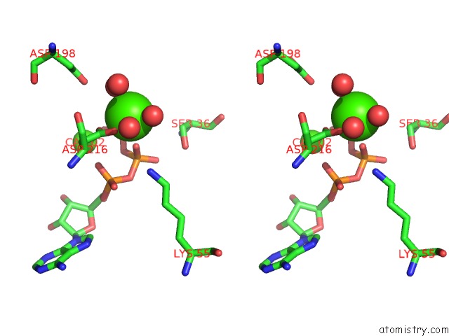
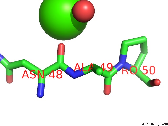
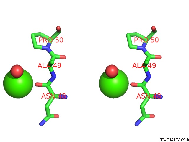
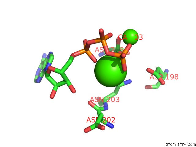
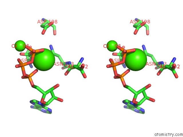
Calcium binding site 2 out of 12 in 4ej7

Go back to Calcium Binding Sites List in 4ej7
Calcium binding site 2 out of 12 in the Crystal Structure of the Aminoglycoside Phosphotransferase Aph(3')-Ia, Atp-Bound


Mono view


Stereo pair view

A full contact list of Calcium with other atoms in the Ca binding site number 2 of Crystal Structure of the Aminoglycoside Phosphotransferase Aph(3')-Ia, Atp-Bound within 5.0Å range:
probe atom residue distance (Å) B Occ
A:Ca403

b:33.2
occ:1.00
O2G A:ATP401 2.2 59.4 1.0
O2B A:ATP401 2.2 24.5 1.0
O A:HOH598 2.3 33.3 1.0
OD2 A:ASP216 2.4 20.4 1.0
O A:HOH599 2.4 29.2 1.0
OD1 A:ASP216 2.5 23.1 1.0
CG A:ASP216 2.7 20.3 1.0
PB A:ATP401 3.5 27.2 1.0
PG A:ATP401 3.5 65.0 1.0
O3B A:ATP401 3.8 48.0 1.0
O A:HOH524 4.0 27.4 1.0
CA A:CA402 4.1 27.9 1.0
OD1 A:ASP198 4.3 26.5 1.0
CB A:ASP216 4.3 15.9 1.0
NZ A:LYS55 4.3 22.0 1.0
O1A A:ATP401 4.3 27.4 1.0
CB A:SER36 4.3 32.9 1.0
O1G A:ATP401 4.3 68.8 1.0
O1B A:ATP401 4.5 29.8 1.0
O3A A:ATP401 4.6 43.9 1.0
OG A:SER36 4.6 34.4 1.0
O3G A:ATP401 4.6 66.6 1.0
PA A:ATP401 4.8 31.1 1.0
O2A A:ATP401 5.0 33.9 1.0

Calcium binding site 3 out of 12 in 4ej7

Go back to Calcium Binding Sites List in 4ej7
Calcium binding site 3 out of 12 in the Crystal Structure of the Aminoglycoside Phosphotransferase Aph(3')-Ia, Atp-Bound


Mono view


Stereo pair view

A full contact list of Calcium with other atoms in the Ca binding site number 3 of Crystal Structure of the Aminoglycoside Phosphotransferase Aph(3')-Ia, Atp-Bound within 5.0Å range:
probe atom residue distance (Å) B Occ
A:Ca404

b:48.4
occ:1.00
O A:HOH538 2.3 31.9 1.0
O A:HOH537 2.4 18.9 1.0
O A:ASP165 2.4 42.4 1.0
OE1 A:GLU269 2.4 26.4 1.0
OD1 A:ASP165 3.5 55.5 1.0
C A:ASP165 3.5 34.3 1.0
O A:HOH536 3.6 38.9 1.0
CD A:GLU269 3.6 25.0 1.0
OD1 A:ASP167 3.7 37.9 1.0
CG A:ASP165 3.9 51.3 1.0
O A:HOH588 4.0 41.3 1.0
CA A:GLU269 4.2 28.1 1.0
CA A:PHE166 4.2 30.7 1.0
N A:PHE166 4.2 31.5 1.0
CB A:ASP165 4.3 43.1 1.0
O A:HOH534 4.3 25.5 1.0
OE2 A:GLU269 4.3 25.7 1.0
C A:PHE166 4.4 31.0 1.0
N A:ASP167 4.4 28.4 1.0
OD2 A:ASP165 4.5 50.6 1.0
CA A:ASP165 4.5 37.6 1.0
O A:ASP268 4.5 23.8 1.0
CG A:ASP167 4.5 36.9 1.0
CG A:GLU269 4.5 24.6 1.0
OD2 A:ASP268 4.6 30.4 1.0
N A:GLU269 4.7 27.9 1.0
CB A:GLU269 4.7 24.1 1.0
C A:ASP268 4.9 27.5 1.0
OD2 A:ASP167 4.9 32.5 1.0

Calcium binding site 4 out of 12 in 4ej7

Go back to Calcium Binding Sites List in 4ej7
Calcium binding site 4 out of 12 in the Crystal Structure of the Aminoglycoside Phosphotransferase Aph(3')-Ia, Atp-Bound


Mono view


Stereo pair view

A full contact list of Calcium with other atoms in the Ca binding site number 4 of Crystal Structure of the Aminoglycoside Phosphotransferase Aph(3')-Ia, Atp-Bound within 5.0Å range:
probe atom residue distance (Å) B Occ
A:Ca405

b:38.7
occ:1.00
O A:ASP31 2.4 28.8 1.0
O A:HOH507 2.4 23.7 1.0
O A:HOH587 2.4 25.9 1.0
O A:HOH603 2.4 44.5 1.0
O A:HOH514 2.4 50.1 1.0
O A:HOH602 2.4 27.1 1.0
O A:HOH508 3.1 31.8 1.0
C A:ASP31 3.5 29.2 1.0
OD2 A:ASP31 4.2 30.2 1.0
CA A:ASN32 4.3 35.4 1.0
N A:ASN32 4.3 31.5 1.0
O A:HOH513 4.4 17.8 1.0
CG A:ARG30 4.4 35.6 1.0
CA A:ASP31 4.5 30.0 1.0
O A:GLY34 4.5 29.5 1.0
N A:ILE40 4.6 29.8 1.0
O A:ALA38 4.6 27.6 1.0
N A:ASP31 4.7 27.4 1.0
CB A:ASP31 4.7 27.9 1.0
O A:ILE40 4.8 24.4 1.0
CD A:ARG30 4.8 38.0 1.0
CG A:ASP31 4.8 27.7 1.0
OD1 A:ASN32 4.8 35.0 1.0
C A:ASN32 4.9 36.5 1.0

Calcium binding site 5 out of 12 in 4ej7

Go back to Calcium Binding Sites List in 4ej7
Calcium binding site 5 out of 12 in the Crystal Structure of the Aminoglycoside Phosphotransferase Aph(3')-Ia, Atp-Bound


Mono view


Stereo pair view

A full contact list of Calcium with other atoms in the Ca binding site number 5 of Crystal Structure of the Aminoglycoside Phosphotransferase Aph(3')-Ia, Atp-Bound within 5.0Å range:
probe atom residue distance (Å) B Occ
B:Ca402

b:36.4
occ:1.00
O1A B:ATP401 2.2 33.2 1.0
O B:HOH579 2.4 40.8 1.0
OD2 B:ASP216 2.4 26.3 1.0
O B:HOH567 2.4 33.4 1.0
OD1 B:ASN203 2.4 35.8 1.0
O2G B:ATP401 2.6 89.8 1.0
O3B B:ATP401 3.1 48.9 1.0
PG B:ATP401 3.3 86.6 1.0
CG B:ASN203 3.4 35.0 1.0
CG B:ASP216 3.4 27.1 1.0
O3G B:ATP401 3.5 84.1 1.0
ND2 B:ASN203 3.7 33.5 1.0
PA B:ATP401 3.7 31.2 1.0
CB B:ASP216 3.8 25.8 1.0
O1B B:ATP401 4.0 46.7 1.0
PB B:ATP401 4.1 39.9 1.0
CA B:CA403 4.2 37.1 1.0
O3A B:ATP401 4.3 65.3 1.0
O5' B:ATP401 4.4 50.7 1.0
C5' B:ATP401 4.4 47.7 1.0
OD1 B:ASP216 4.5 27.8 1.0
OD1 B:ASP198 4.6 46.0 1.0
O2A B:ATP401 4.7 34.6 1.0
O B:ASP202 4.7 38.5 1.0
O1G B:ATP401 4.7 85.4 1.0
CB B:ASN203 4.8 30.3 1.0
C B:ASP202 5.0 38.3 1.0
O B:HOH574 5.0 40.1 1.0

Calcium binding site 6 out of 12 in 4ej7

Go back to Calcium Binding Sites List in 4ej7
Calcium binding site 6 out of 12 in the Crystal Structure of the Aminoglycoside Phosphotransferase Aph(3')-Ia, Atp-Bound


Mono view


Stereo pair view

A full contact list of Calcium with other atoms in the Ca binding site number 6 of Crystal Structure of the Aminoglycoside Phosphotransferase Aph(3')-Ia, Atp-Bound within 5.0Å range:
probe atom residue distance (Å) B Occ
B:Ca403

b:37.1
occ:1.00
O3G B:ATP401 2.3 84.1 1.0
O1B B:ATP401 2.3 46.7 1.0
O B:HOH559 2.3 31.0 1.0
O B:HOH560 2.4 32.5 1.0
OD2 B:ASP216 2.4 26.3 1.0
OD1 B:ASP216 2.5 27.8 1.0
CG B:ASP216 2.8 27.1 1.0
PB B:ATP401 3.6 39.9 1.0
PG B:ATP401 3.7 86.6 1.0
O B:HOH511 3.8 43.2 1.0
O3B B:ATP401 3.9 48.9 1.0
OD1 B:ASP198 4.2 46.0 1.0
CA B:CA402 4.2 36.4 1.0
CB B:ASP216 4.3 25.8 1.0
O B:HOH524 4.3 28.4 1.0
CB B:SER36 4.3 58.0 1.0
O1A B:ATP401 4.4 33.2 1.0
O2B B:ATP401 4.5 38.0 1.0
O2G B:ATP401 4.5 89.8 1.0
NZ B:LYS55 4.6 31.2 1.0
O1G B:ATP401 4.7 85.4 1.0
O3A B:ATP401 4.7 65.3 1.0
OG B:SER36 4.8 58.0 1.0
PA B:ATP401 4.9 31.2 1.0

Calcium binding site 7 out of 12 in 4ej7

Go back to Calcium Binding Sites List in 4ej7
Calcium binding site 7 out of 12 in the Crystal Structure of the Aminoglycoside Phosphotransferase Aph(3')-Ia, Atp-Bound


Mono view


Stereo pair view

A full contact list of Calcium with other atoms in the Ca binding site number 7 of Crystal Structure of the Aminoglycoside Phosphotransferase Aph(3')-Ia, Atp-Bound within 5.0Å range:
probe atom residue distance (Å) B Occ
B:Ca404

b:67.0
occ:1.00
O B:HOH583 2.4 41.0 1.0
OE1 B:GLU269 2.4 42.9 1.0
O B:ASP165 2.4 61.1 1.0
C B:ASP165 3.6 61.4 1.0
CD B:GLU269 3.6 40.8 1.0
CA B:PHE166 4.1 51.4 1.0
OD1 B:ASP167 4.1 61.1 1.0
OD1 B:ASP165 4.2 73.2 1.0
CA B:GLU269 4.2 39.4 1.0
N B:PHE166 4.2 54.8 1.0
C B:PHE166 4.3 55.2 1.0
CG B:ASP165 4.3 72.4 1.0
N B:ASP167 4.3 53.8 1.0
OE2 B:GLU269 4.4 39.3 1.0
CB B:ASP165 4.5 70.7 1.0
CG B:GLU269 4.6 39.5 1.0
CB B:GLU269 4.7 38.1 1.0
CA B:ASP165 4.7 68.7 1.0
CG B:ASP167 4.7 57.1 1.0
O B:ASP268 4.7 44.5 1.0
O B:GLU269 4.8 36.5 1.0
OD2 B:ASP165 4.8 72.5 1.0
N B:GLU269 4.9 40.0 1.0
C B:GLU269 5.0 38.8 1.0
CD1 B:PHE166 5.0 40.2 1.0
O B:PHE166 5.0 58.6 1.0

Calcium binding site 8 out of 12 in 4ej7

Go back to Calcium Binding Sites List in 4ej7
Calcium binding site 8 out of 12 in the Crystal Structure of the Aminoglycoside Phosphotransferase Aph(3')-Ia, Atp-Bound


Mono view


Stereo pair view

A full contact list of Calcium with other atoms in the Ca binding site number 8 of Crystal Structure of the Aminoglycoside Phosphotransferase Aph(3')-Ia, Atp-Bound within 5.0Å range:
probe atom residue distance (Å) B Occ
B:Ca405

b:77.3
occ:1.00
O B:HOH561 2.4 59.2 1.0
O B:ASN48 2.4 45.3 1.0
C B:ASN48 3.6 41.2 1.0
CB B:ASN48 4.5 36.9 1.0
CA B:ASN48 4.5 40.6 1.0
N B:ALA49 4.5 37.1 1.0
CA B:ALA49 4.6 33.9 1.0
CD B:PRO50 4.9 41.1 1.0

Calcium binding site 9 out of 12 in 4ej7

Go back to Calcium Binding Sites List in 4ej7
Calcium binding site 9 out of 12 in the Crystal Structure of the Aminoglycoside Phosphotransferase Aph(3')-Ia, Atp-Bound


Mono view


Stereo pair view

A full contact list of Calcium with other atoms in the Ca binding site number 9 of Crystal Structure of the Aminoglycoside Phosphotransferase Aph(3')-Ia, Atp-Bound within 5.0Å range:
probe atom residue distance (Å) B Occ
B:Ca406

b:85.7
occ:1.00
O B:ASP31 2.3 88.6 1.0
O B:HOH580 2.4 61.7 1.0
O B:HOH568 2.4 62.3 1.0
C B:ASP31 3.2 87.7 1.0
N B:ASP31 3.6 87.0 1.0
CA B:ASP31 3.8 86.6 1.0
O B:ILE40 4.0 52.2 1.0
CB B:ASP31 4.1 84.6 1.0
N B:ASN32 4.3 86.7 1.0
N B:ILE40 4.3 44.5 1.0
ND2 B:ASN32 4.4 82.7 1.0
C B:ARG30 4.4 86.1 1.0
CB B:ARG30 4.5 90.5 1.0
CA B:ASN32 4.6 84.3 1.0
OD2 B:ASP31 4.8 82.3 1.0
CA B:ARG30 4.8 84.7 1.0
C B:ILE40 4.9 49.1 1.0
CG B:ASP31 5.0 83.8 1.0
CA B:ILE40 5.0 43.8 1.0

Calcium binding site 10 out of 12 in 4ej7

Go back to Calcium Binding Sites List in 4ej7
Calcium binding site 10 out of 12 in the Crystal Structure of the Aminoglycoside Phosphotransferase Aph(3')-Ia, Atp-Bound


Mono view


Stereo pair view

A full contact list of Calcium with other atoms in the Ca binding site number 10 of Crystal Structure of the Aminoglycoside Phosphotransferase Aph(3')-Ia, Atp-Bound within 5.0Å range:
probe atom residue distance (Å) B Occ
C:Ca402

b:32.3
occ:1.00
O1A C:ATP401 2.1 29.1 1.0
O2G C:ATP401 2.3 72.8 1.0
OD1 C:ASN203 2.4 32.1 1.0
OD1 C:ASP216 2.5 28.9 1.0
O3B C:ATP401 2.8 58.5 1.0
PG C:ATP401 3.1 71.8 1.0
CG C:ASP216 3.4 22.6 1.0
CG C:ASN203 3.4 31.0 1.0
PA C:ATP401 3.5 32.1 1.0
CB C:ASP216 3.7 21.3 1.0
O3G C:ATP401 3.7 70.1 1.0
ND2 C:ASN203 3.8 33.3 1.0
PB C:ATP401 3.9 28.8 1.0
O1B C:ATP401 4.0 32.6 1.0
O3A C:ATP401 4.0 25.8 1.0
CA C:CA403 4.3 39.0 1.0
OD2 C:ASP216 4.4 25.7 1.0
O1G C:ATP401 4.4 71.1 1.0
OD1 C:ASP198 4.4 38.6 1.0
O5' C:ATP401 4.5 26.8 1.0
O2A C:ATP401 4.5 34.6 1.0
O C:ASP202 4.5 35.4 1.0
O3' C:ATP401 4.6 36.1 1.0
C5' C:ATP401 4.7 24.1 1.0
CB C:ASN203 4.7 30.2 1.0
C C:ASP202 4.8 36.6 1.0
C3' C:ATP401 4.9 30.8 1.0
CB C:ASP202 5.0 45.8 1.0

Reference:

P.J.Stogios, P.Spanogiannopoulos, E.Evdokimova, O.Egorova, T.Shakya, N.Todorovic, A.Capretta, G.D.Wright, A.Savchenko. Structure-Guided Optimization of Protein Kinase Inhibitors Reverses Aminoglycoside Antibiotic Resistance. Biochem.J. V. 454 191 2013.
ISSN: ISSN 0264-6021
PubMed: 23758273
DOI: 10.1042/BJ20130317
Page generated: Sat Dec 12 04:45:04 2020

Last articles

Zn in 8WB0
Zn in 8WAX
Zn in 8WAU
Zn in 8WAZ
Zn in 8WAY
Zn in 8WAV
Zn in 8WAW
Zn in 8WAT
Zn in 8W7M
Zn in 8WD3
© Copyright 2008-2020 by atomistry.com
Home   |    Site Map   |    Copyright   |    Contact us   |    Privacy