Calcium »
PDB 4ecg-4enz »
4emt »
Calcium in PDB 4emt: Crystal Structure of Human Sting Bound to C-Di-Gmp
Protein crystallography data
The structure of Crystal Structure of Human Sting Bound to C-Di-Gmp, PDB code: 4emt
was solved by
P.Li,
with X-Ray Crystallography technique. A brief refinement statistics is given in the table below:
| Resolution Low / High (Å) | 48.25 / 1.50 |
| Space group | C 2 2 21 |
| Cell size a, b, c (Å), α, β, γ (°) | 69.797, 78.215, 128.222, 90.00, 90.00, 90.00 |
| R / Rfree (%) | 18.8 / 21 |
Calcium Binding Sites:
The binding sites of Calcium atom in the Crystal Structure of Human Sting Bound to C-Di-Gmp
(pdb code 4emt). This binding sites where shown within
5.0 Angstroms radius around Calcium atom.
In total only one binding site of Calcium was determined in the Crystal Structure of Human Sting Bound to C-Di-Gmp, PDB code: 4emt:
In total only one binding site of Calcium was determined in the Crystal Structure of Human Sting Bound to C-Di-Gmp, PDB code: 4emt:
Calcium binding site 1 out of 1 in 4emt
Go back to
Calcium binding site 1 out
of 1 in the Crystal Structure of Human Sting Bound to C-Di-Gmp
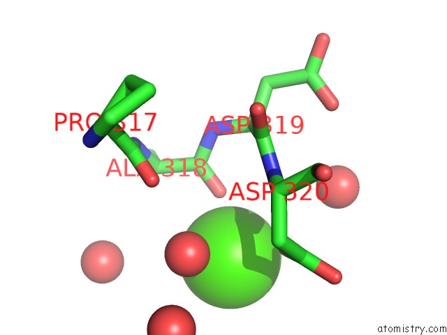
Mono view
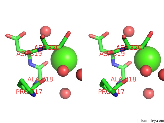
Stereo pair view
Mono view
Stereo pair view
A full contact list of Calcium with other atoms in the Ca binding
site number 1 of Crystal Structure of Human Sting Bound to C-Di-Gmp within 5.0Å range:
|
Reference:
C.Shu,
G.Yi,
T.Watts,
C.C.Kao,
P.Li.
Structure of Sting Bound to Cyclic Di-Gmp Reveals the Mechanism of Cyclic Dinucleotide Recognition By the Immune System. Nat.Struct.Mol.Biol. V. 19 722 2012.
ISSN: ISSN 1545-9993
PubMed: 22728658
DOI: 10.1038/NSMB.2331
Page generated: Tue Jul 8 19:49:55 2025
ISSN: ISSN 1545-9993
PubMed: 22728658
DOI: 10.1038/NSMB.2331
Last articles
Cl in 8C3LCl in 8C2F
Cl in 8C25
Cl in 8C3F
Cl in 8C2E
Cl in 8C1P
Cl in 8C26
Cl in 8C1K
Cl in 8C1H
Cl in 8C14