Calcium »
PDB 4eoy-4fb1 »
4evh »
Calcium in PDB 4evh: Crystal Structure of Calcium-Bound Alpha-1 Giardin
Protein crystallography data
The structure of Crystal Structure of Calcium-Bound Alpha-1 Giardin, PDB code: 4evh
was solved by
S.Weeratunga,
A.Hofmann,
with X-Ray Crystallography technique. A brief refinement statistics is given in the table below:
| Resolution Low / High (Å) | 24.83 / 2.60 |
| Space group | P 21 21 21 |
| Cell size a, b, c (Å), α, β, γ (°) | 51.309, 58.363, 98.762, 90.00, 90.00, 90.00 |
| R / Rfree (%) | 21.3 / 30.4 |
Calcium Binding Sites:
The binding sites of Calcium atom in the Crystal Structure of Calcium-Bound Alpha-1 Giardin
(pdb code 4evh). This binding sites where shown within
5.0 Angstroms radius around Calcium atom.
In total 2 binding sites of Calcium where determined in the Crystal Structure of Calcium-Bound Alpha-1 Giardin, PDB code: 4evh:
Jump to Calcium binding site number: 1; 2;
In total 2 binding sites of Calcium where determined in the Crystal Structure of Calcium-Bound Alpha-1 Giardin, PDB code: 4evh:
Jump to Calcium binding site number: 1; 2;
Calcium binding site 1 out of 2 in 4evh
Go back to
Calcium binding site 1 out
of 2 in the Crystal Structure of Calcium-Bound Alpha-1 Giardin
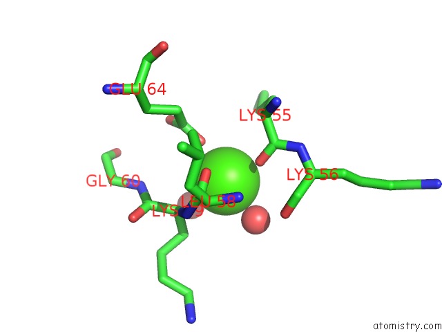
Mono view
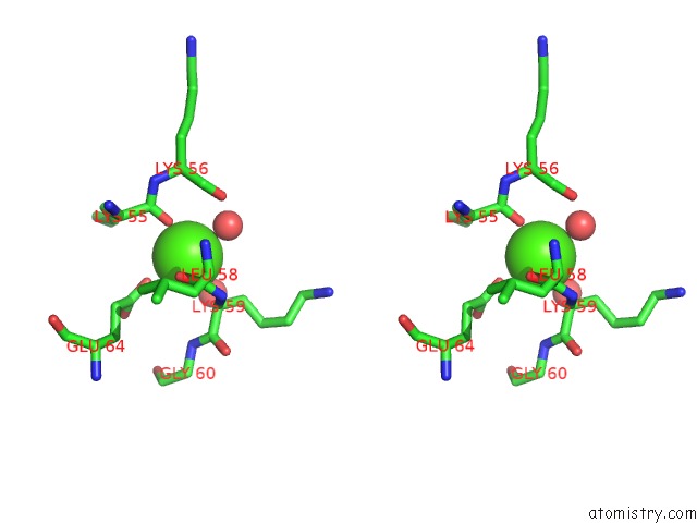
Stereo pair view
Mono view
Stereo pair view
A full contact list of Calcium with other atoms in the Ca binding
site number 1 of Crystal Structure of Calcium-Bound Alpha-1 Giardin within 5.0Å range:
|
Calcium binding site 2 out of 2 in 4evh
Go back to
Calcium binding site 2 out
of 2 in the Crystal Structure of Calcium-Bound Alpha-1 Giardin
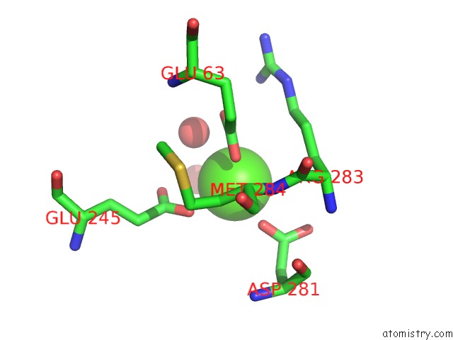
Mono view
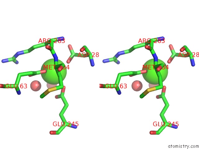
Stereo pair view
Mono view
Stereo pair view
A full contact list of Calcium with other atoms in the Ca binding
site number 2 of Crystal Structure of Calcium-Bound Alpha-1 Giardin within 5.0Å range:
|
Reference:
S.K.Weeratunga,
A.Osman,
N.-J.Hu,
C.K.Wang,
L.Mason,
S.Svard,
G.Hope,
M.K.Jones,
A.Hofmann.
Alpha-1 Giardin Is An Annexin with Highly Unusual Calcium-Regulated Mechanisms J.Mol.Biol. V. 423 169 2012.
ISSN: ISSN 0022-2836
PubMed: 22796298
DOI: 10.1016/J.JMB.2012.06.041
Page generated: Tue Jul 8 19:52:38 2025
ISSN: ISSN 0022-2836
PubMed: 22796298
DOI: 10.1016/J.JMB.2012.06.041
Last articles
Cl in 5QF7Cl in 5QED
Cl in 5QFA
Cl in 5QEW
Cl in 5QEJ
Cl in 5QEV
Cl in 5QES
Cl in 5QEU
Cl in 5QDH
Cl in 5QEB