Calcium »
PDB 4fbt-4fs8 »
4fef »
Calcium in PDB 4fef: The Crystal Structures of Several Mutants of Pleurotus Eryngii Versatile Peroxidase
Enzymatic activity of The Crystal Structures of Several Mutants of Pleurotus Eryngii Versatile Peroxidase
All present enzymatic activity of The Crystal Structures of Several Mutants of Pleurotus Eryngii Versatile Peroxidase:
1.11.1.16;
1.11.1.16;
Protein crystallography data
The structure of The Crystal Structures of Several Mutants of Pleurotus Eryngii Versatile Peroxidase, PDB code: 4fef
was solved by
M.J.Mate,
A.Romero,
F.J.Ruiz-Duenas,
A.T.Martinez,
with X-Ray Crystallography technique. A brief refinement statistics is given in the table below:
| Resolution Low / High (Å) | 68.81 / 2.00 |
| Space group | I 41 |
| Cell size a, b, c (Å), α, β, γ (°) | 96.584, 96.584, 98.065, 90.00, 90.00, 90.00 |
| R / Rfree (%) | 13.8 / 17.6 |
Other elements in 4fef:
The structure of The Crystal Structures of Several Mutants of Pleurotus Eryngii Versatile Peroxidase also contains other interesting chemical elements:
| Iron | (Fe) | 1 atom |
Calcium Binding Sites:
The binding sites of Calcium atom in the The Crystal Structures of Several Mutants of Pleurotus Eryngii Versatile Peroxidase
(pdb code 4fef). This binding sites where shown within
5.0 Angstroms radius around Calcium atom.
In total 2 binding sites of Calcium where determined in the The Crystal Structures of Several Mutants of Pleurotus Eryngii Versatile Peroxidase, PDB code: 4fef:
Jump to Calcium binding site number: 1; 2;
In total 2 binding sites of Calcium where determined in the The Crystal Structures of Several Mutants of Pleurotus Eryngii Versatile Peroxidase, PDB code: 4fef:
Jump to Calcium binding site number: 1; 2;
Calcium binding site 1 out of 2 in 4fef
Go back to
Calcium binding site 1 out
of 2 in the The Crystal Structures of Several Mutants of Pleurotus Eryngii Versatile Peroxidase
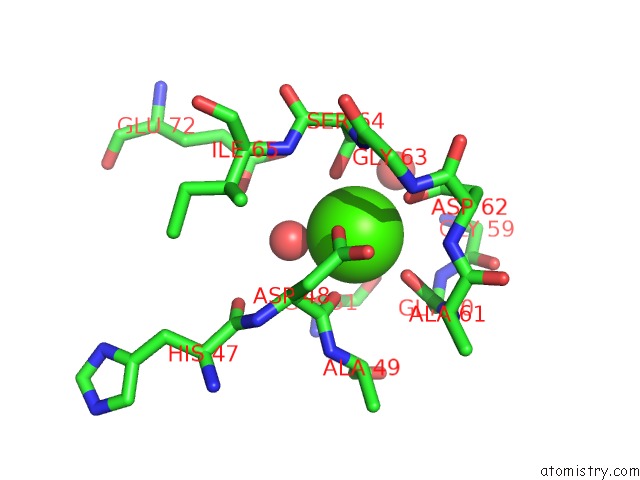
Mono view
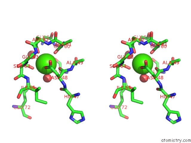
Stereo pair view
Mono view
Stereo pair view
A full contact list of Calcium with other atoms in the Ca binding
site number 1 of The Crystal Structures of Several Mutants of Pleurotus Eryngii Versatile Peroxidase within 5.0Å range:
|
Calcium binding site 2 out of 2 in 4fef
Go back to
Calcium binding site 2 out
of 2 in the The Crystal Structures of Several Mutants of Pleurotus Eryngii Versatile Peroxidase
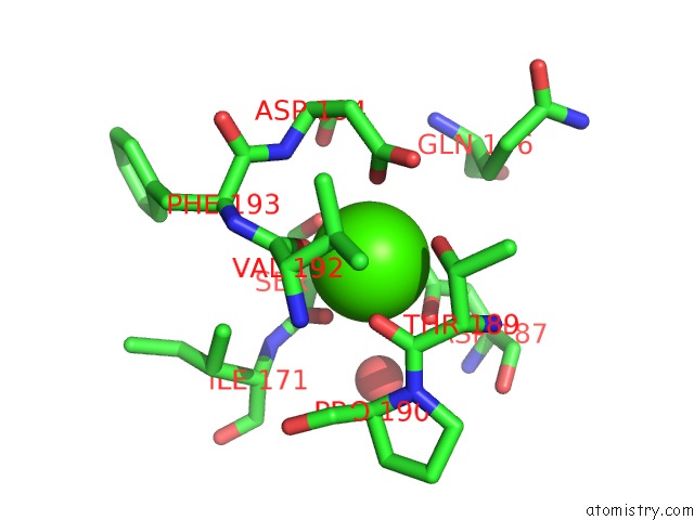
Mono view
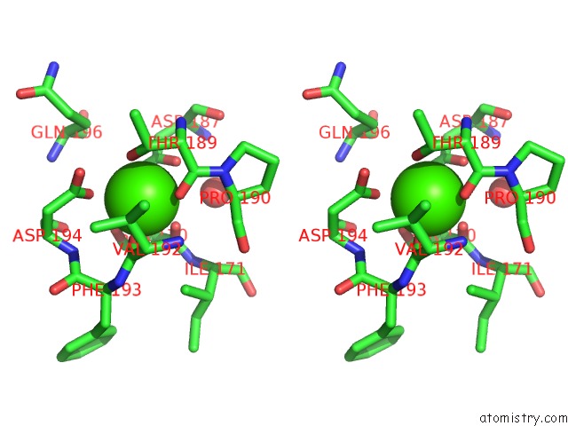
Stereo pair view
Mono view
Stereo pair view
A full contact list of Calcium with other atoms in the Ca binding
site number 2 of The Crystal Structures of Several Mutants of Pleurotus Eryngii Versatile Peroxidase within 5.0Å range:
|
Reference:
M.Morales,
M.J.Mate,
A.Romero,
M.J.Martinez,
A.T.Martinez,
F.J.Ruiz-Duenas.
Two Oxidation Sites For Low Redox Potential Substrates: A Directed Mutagenesis, Kinetic, and Crystallographic Study on Pleurotus Eryngii Versatile Peroxidase. J.Biol.Chem. V. 287 41053 2012.
ISSN: ISSN 0021-9258
PubMed: 23071108
DOI: 10.1074/JBC.M112.405548
Page generated: Sun Jul 14 00:17:49 2024
ISSN: ISSN 0021-9258
PubMed: 23071108
DOI: 10.1074/JBC.M112.405548
Last articles
Zn in 9J0NZn in 9J0O
Zn in 9J0P
Zn in 9FJX
Zn in 9EKB
Zn in 9C0F
Zn in 9CAH
Zn in 9CH0
Zn in 9CH3
Zn in 9CH1