Calcium »
PDB 4fbt-4fs8 »
4fqo »
Calcium in PDB 4fqo: Crystal Structure of Calcium-Loaded S100B Bound to SBI4211
Protein crystallography data
The structure of Crystal Structure of Calcium-Loaded S100B Bound to SBI4211, PDB code: 4fqo
was solved by
L.E.Mcknight,
E.P.Raman,
P.Bezawada,
S.Kudrimoti,
P.T.Wilder,
K.G.Hartman,
E.A.Toth,
A.Coop,
A.D.Mackerrell,
D.J.Weber,
with X-Ray Crystallography technique. A brief refinement statistics is given in the table below:
| Resolution Low / High (Å) | 44.60 / 1.65 |
| Space group | P 41 21 2 |
| Cell size a, b, c (Å), α, β, γ (°) | 63.079, 63.079, 48.998, 90.00, 90.00, 90.00 |
| R / Rfree (%) | 19.9 / 21.4 |
Calcium Binding Sites:
The binding sites of Calcium atom in the Crystal Structure of Calcium-Loaded S100B Bound to SBI4211
(pdb code 4fqo). This binding sites where shown within
5.0 Angstroms radius around Calcium atom.
In total 2 binding sites of Calcium where determined in the Crystal Structure of Calcium-Loaded S100B Bound to SBI4211, PDB code: 4fqo:
Jump to Calcium binding site number: 1; 2;
In total 2 binding sites of Calcium where determined in the Crystal Structure of Calcium-Loaded S100B Bound to SBI4211, PDB code: 4fqo:
Jump to Calcium binding site number: 1; 2;
Calcium binding site 1 out of 2 in 4fqo
Go back to
Calcium binding site 1 out
of 2 in the Crystal Structure of Calcium-Loaded S100B Bound to SBI4211

Mono view
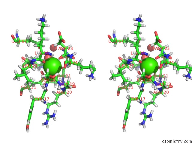
Stereo pair view
Mono view
Stereo pair view
A full contact list of Calcium with other atoms in the Ca binding
site number 1 of Crystal Structure of Calcium-Loaded S100B Bound to SBI4211 within 5.0Å range:
|
Calcium binding site 2 out of 2 in 4fqo
Go back to
Calcium binding site 2 out
of 2 in the Crystal Structure of Calcium-Loaded S100B Bound to SBI4211

Mono view

Stereo pair view
Mono view
Stereo pair view
A full contact list of Calcium with other atoms in the Ca binding
site number 2 of Crystal Structure of Calcium-Loaded S100B Bound to SBI4211 within 5.0Å range:
|
Reference:
L.E.Mcknight,
E.P.Raman,
P.Bezawada,
S.Kudrimoti,
P.T.Wilder,
K.G.Hartman,
R.Godoy-Ruiz,
E.A.Toth,
A.Coop,
A.D.Mackerell,
D.J.Weber.
Structure-Based Discovery of A Novel Pentamidine-Related Inhibitor of the Calcium-Binding Protein S100B. Acs Med Chem Lett V. 3 975 2012.
ISSN: ISSN 1948-5875
PubMed: 23264854
DOI: 10.1021/ML300166S
Page generated: Sun Jul 14 00:26:49 2024
ISSN: ISSN 1948-5875
PubMed: 23264854
DOI: 10.1021/ML300166S
Last articles
Zn in 9MJ5Zn in 9HNW
Zn in 9G0L
Zn in 9FNE
Zn in 9DZN
Zn in 9E0I
Zn in 9D32
Zn in 9DAK
Zn in 8ZXC
Zn in 8ZUF