Calcium »
PDB 4fsd-4gg1 »
4gc6 »
Calcium in PDB 4gc6: Crystal Structure of DPO4 in Complex with N-Mc-Damp Opposite Dt
Enzymatic activity of Crystal Structure of DPO4 in Complex with N-Mc-Damp Opposite Dt
All present enzymatic activity of Crystal Structure of DPO4 in Complex with N-Mc-Damp Opposite Dt:
2.7.7.7;
2.7.7.7;
Protein crystallography data
The structure of Crystal Structure of DPO4 in Complex with N-Mc-Damp Opposite Dt, PDB code: 4gc6
was solved by
R.L.Eoff,
A.Ketkar,
S.Banerjee,
M.K.Zafar,
with X-Ray Crystallography technique. A brief refinement statistics is given in the table below:
| Resolution Low / High (Å) | 45.79 / 2.90 |
| Space group | P 21 21 2 |
| Cell size a, b, c (Å), α, β, γ (°) | 93.641, 103.357, 52.491, 90.00, 90.00, 90.00 |
| R / Rfree (%) | 20.2 / 27.6 |
Calcium Binding Sites:
The binding sites of Calcium atom in the Crystal Structure of DPO4 in Complex with N-Mc-Damp Opposite Dt
(pdb code 4gc6). This binding sites where shown within
5.0 Angstroms radius around Calcium atom.
In total 4 binding sites of Calcium where determined in the Crystal Structure of DPO4 in Complex with N-Mc-Damp Opposite Dt, PDB code: 4gc6:
Jump to Calcium binding site number: 1; 2; 3; 4;
In total 4 binding sites of Calcium where determined in the Crystal Structure of DPO4 in Complex with N-Mc-Damp Opposite Dt, PDB code: 4gc6:
Jump to Calcium binding site number: 1; 2; 3; 4;
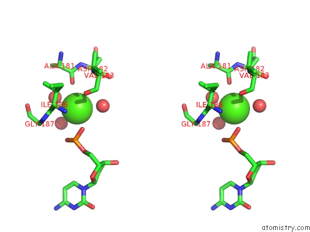
Calcium binding site 1 out of 4 in 4gc6
Go back to
Calcium binding site 1 out
of 4 in the Crystal Structure of DPO4 in Complex with N-Mc-Damp Opposite Dt
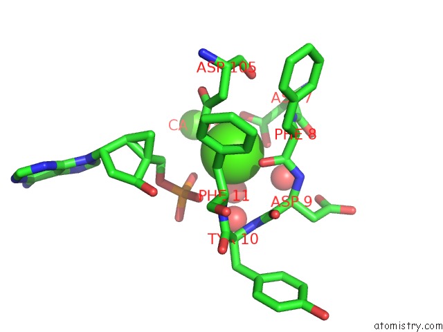
Mono view
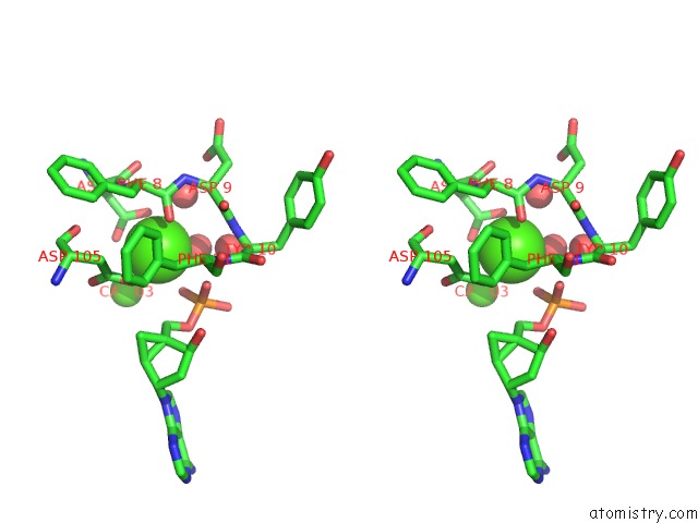
Stereo pair view
Mono view
Stereo pair view
A full contact list of Calcium with other atoms in the Ca binding
site number 1 of Crystal Structure of DPO4 in Complex with N-Mc-Damp Opposite Dt within 5.0Å range:
|
Calcium binding site 2 out of 4 in 4gc6
Go back to
Calcium binding site 2 out
of 4 in the Crystal Structure of DPO4 in Complex with N-Mc-Damp Opposite Dt
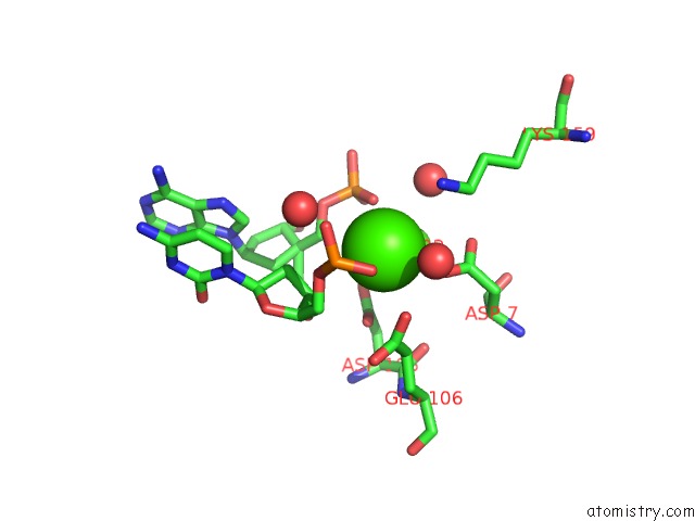
Mono view

Stereo pair view
Mono view
Stereo pair view
A full contact list of Calcium with other atoms in the Ca binding
site number 2 of Crystal Structure of DPO4 in Complex with N-Mc-Damp Opposite Dt within 5.0Å range:
|
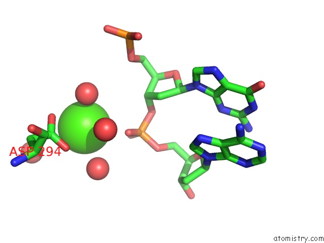
Calcium binding site 3 out of 4 in 4gc6
Go back to
Calcium binding site 3 out
of 4 in the Crystal Structure of DPO4 in Complex with N-Mc-Damp Opposite Dt
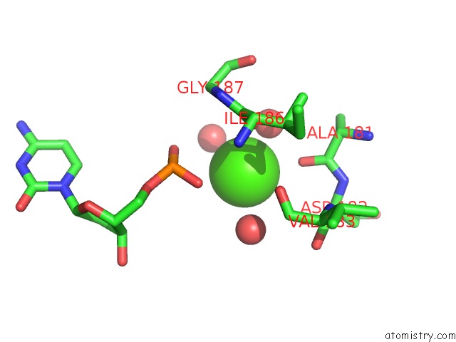
Mono view
Stereo pair view
Mono view
Stereo pair view
A full contact list of Calcium with other atoms in the Ca binding
site number 3 of Crystal Structure of DPO4 in Complex with N-Mc-Damp Opposite Dt within 5.0Å range:
|
Calcium binding site 4 out of 4 in 4gc6
Go back to
Calcium binding site 4 out
of 4 in the Crystal Structure of DPO4 in Complex with N-Mc-Damp Opposite Dt
Mono view
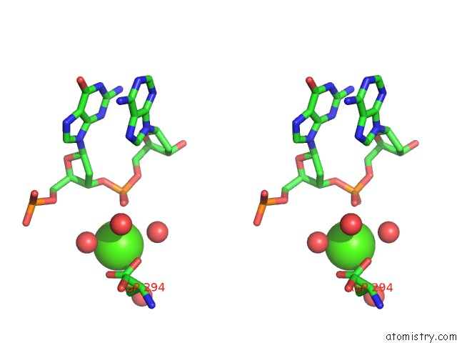
Stereo pair view
Mono view
Stereo pair view
A full contact list of Calcium with other atoms in the Ca binding
site number 4 of Crystal Structure of DPO4 in Complex with N-Mc-Damp Opposite Dt within 5.0Å range:
|
Reference:
A.Ketkar,
M.K.Zafar,
S.Banerjee,
V.E.Marquez,
M.Egli,
R.L.Eoff.
Differential Furanose Selection in the Active Sites of Archaeal Dna Polymerases Probed By Fixed-Conformation Nucleotide Analogues. Biochemistry V. 51 9234 2012.
ISSN: ISSN 0006-2960
PubMed: 23050956
DOI: 10.1021/BI301043K
Page generated: Tue Jul 8 20:14:55 2025
ISSN: ISSN 0006-2960
PubMed: 23050956
DOI: 10.1021/BI301043K
Last articles
Ca in 7NS9Ca in 7NRK
Ca in 7NPG
Ca in 7NPB
Ca in 7NN9
Ca in 7NMX
Ca in 7NN2
Ca in 7NIT
Ca in 7NMH
Ca in 7NM3