Calcium »
PDB 4ggb-4gpe »
4gh8 »
Calcium in PDB 4gh8: Crystal Structure of A 'Humanized' E. Coli Dihydrofolate Reductase
Enzymatic activity of Crystal Structure of A 'Humanized' E. Coli Dihydrofolate Reductase
All present enzymatic activity of Crystal Structure of A 'Humanized' E. Coli Dihydrofolate Reductase:
1.5.1.3;
1.5.1.3;
Protein crystallography data
The structure of Crystal Structure of A 'Humanized' E. Coli Dihydrofolate Reductase, PDB code: 4gh8
was solved by
J.B.French,
C.T.Liu,
P.Hanoian,
T.H.Pringle,
S.Hammes-Schiffer,
S.J.Benkovic,
with X-Ray Crystallography technique. A brief refinement statistics is given in the table below:
| Resolution Low / High (Å) | 50.00 / 1.85 |
| Space group | P 1 21 1 |
| Cell size a, b, c (Å), α, β, γ (°) | 52.245, 63.773, 62.439, 90.00, 106.82, 90.00 |
| R / Rfree (%) | 20.3 / 24.5 |
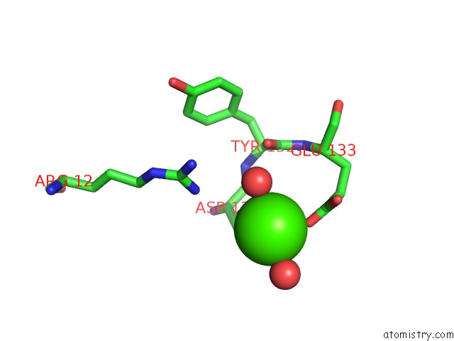
Calcium Binding Sites:
The binding sites of Calcium atom in the Crystal Structure of A 'Humanized' E. Coli Dihydrofolate Reductase
(pdb code 4gh8). This binding sites where shown within
5.0 Angstroms radius around Calcium atom.
In total 3 binding sites of Calcium where determined in the Crystal Structure of A 'Humanized' E. Coli Dihydrofolate Reductase, PDB code: 4gh8:
Jump to Calcium binding site number: 1; 2; 3;
In total 3 binding sites of Calcium where determined in the Crystal Structure of A 'Humanized' E. Coli Dihydrofolate Reductase, PDB code: 4gh8:
Jump to Calcium binding site number: 1; 2; 3;
Calcium binding site 1 out of 3 in 4gh8
Go back to
Calcium binding site 1 out
of 3 in the Crystal Structure of A 'Humanized' E. Coli Dihydrofolate Reductase

Mono view

Stereo pair view
Mono view
Stereo pair view
A full contact list of Calcium with other atoms in the Ca binding
site number 1 of Crystal Structure of A 'Humanized' E. Coli Dihydrofolate Reductase within 5.0Å range:
|
Calcium binding site 2 out of 3 in 4gh8
Go back to
Calcium binding site 2 out
of 3 in the Crystal Structure of A 'Humanized' E. Coli Dihydrofolate Reductase
Mono view

Stereo pair view
Mono view
Stereo pair view
A full contact list of Calcium with other atoms in the Ca binding
site number 2 of Crystal Structure of A 'Humanized' E. Coli Dihydrofolate Reductase within 5.0Å range:
|
Calcium binding site 3 out of 3 in 4gh8
Go back to
Calcium binding site 3 out
of 3 in the Crystal Structure of A 'Humanized' E. Coli Dihydrofolate Reductase

Mono view

Stereo pair view
Mono view
Stereo pair view
A full contact list of Calcium with other atoms in the Ca binding
site number 3 of Crystal Structure of A 'Humanized' E. Coli Dihydrofolate Reductase within 5.0Å range:
|
Reference:
C.T.Liu,
P.Hanoian,
J.B.French,
T.H.Pringle,
S.Hammes-Schiffer,
S.J.Benkovic.
Functional Significance of Evolving Protein Sequence in Dihydrofolate Reductase From Bacteria to Humans. Proc.Natl.Acad.Sci.Usa V. 110 10159 2013.
ISSN: ISSN 0027-8424
PubMed: 23733948
DOI: 10.1073/PNAS.1307130110
Page generated: Sun Jul 14 07:13:12 2024
ISSN: ISSN 0027-8424
PubMed: 23733948
DOI: 10.1073/PNAS.1307130110
Last articles
Zn in 9MJ5Zn in 9HNW
Zn in 9G0L
Zn in 9FNE
Zn in 9DZN
Zn in 9E0I
Zn in 9D32
Zn in 9DAK
Zn in 8ZXC
Zn in 8ZUF