Calcium »
PDB 4ggb-4gpe »
4ghg »
Calcium in PDB 4ghg: Structure of Homoprotocatechuate 2,3-Dioxygenase From B.Fuscum in Complex with Hpca at 1.50 Ang Resolution
Enzymatic activity of Structure of Homoprotocatechuate 2,3-Dioxygenase From B.Fuscum in Complex with Hpca at 1.50 Ang Resolution
All present enzymatic activity of Structure of Homoprotocatechuate 2,3-Dioxygenase From B.Fuscum in Complex with Hpca at 1.50 Ang Resolution:
1.13.11.15;
1.13.11.15;
Protein crystallography data
The structure of Structure of Homoprotocatechuate 2,3-Dioxygenase From B.Fuscum in Complex with Hpca at 1.50 Ang Resolution, PDB code: 4ghg
was solved by
E.G.Kovaleva,
J.D.Lipscomb,
with X-Ray Crystallography technique. A brief refinement statistics is given in the table below:
| Resolution Low / High (Å) | 45.86 / 1.50 |
| Space group | P 21 21 2 |
| Cell size a, b, c (Å), α, β, γ (°) | 110.540, 150.785, 96.276, 90.00, 90.00, 90.00 |
| R / Rfree (%) | 12.7 / 16.1 |
Other elements in 4ghg:
The structure of Structure of Homoprotocatechuate 2,3-Dioxygenase From B.Fuscum in Complex with Hpca at 1.50 Ang Resolution also contains other interesting chemical elements:
| Iron | (Fe) | 4 atoms |
| Chlorine | (Cl) | 3 atoms |
Calcium Binding Sites:
The binding sites of Calcium atom in the Structure of Homoprotocatechuate 2,3-Dioxygenase From B.Fuscum in Complex with Hpca at 1.50 Ang Resolution
(pdb code 4ghg). This binding sites where shown within
5.0 Angstroms radius around Calcium atom.
In total only one binding site of Calcium was determined in the Structure of Homoprotocatechuate 2,3-Dioxygenase From B.Fuscum in Complex with Hpca at 1.50 Ang Resolution, PDB code: 4ghg:
In total only one binding site of Calcium was determined in the Structure of Homoprotocatechuate 2,3-Dioxygenase From B.Fuscum in Complex with Hpca at 1.50 Ang Resolution, PDB code: 4ghg:
Calcium binding site 1 out of 1 in 4ghg
Go back to
Calcium binding site 1 out
of 1 in the Structure of Homoprotocatechuate 2,3-Dioxygenase From B.Fuscum in Complex with Hpca at 1.50 Ang Resolution
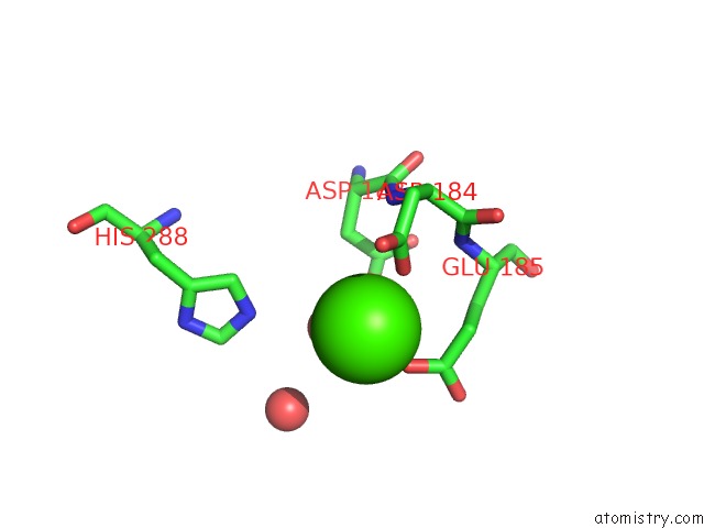
Mono view
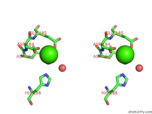
Stereo pair view
Mono view
Stereo pair view
A full contact list of Calcium with other atoms in the Ca binding
site number 1 of Structure of Homoprotocatechuate 2,3-Dioxygenase From B.Fuscum in Complex with Hpca at 1.50 Ang Resolution within 5.0Å range:
|
Reference:
E.G.Kovaleva,
J.D.Lipscomb.
Structural Basis For the Role of Tyrosine 257 of Homoprotocatechuate 2,3-Dioxygenase in Substrate and Oxygen Activation. Biochemistry V. 51 8755 2012.
ISSN: ISSN 0006-2960
PubMed: 23066739
DOI: 10.1021/BI301115C
Page generated: Sun Jul 14 07:13:50 2024
ISSN: ISSN 0006-2960
PubMed: 23066739
DOI: 10.1021/BI301115C
Last articles
Zn in 9MJ5Zn in 9HNW
Zn in 9G0L
Zn in 9FNE
Zn in 9DZN
Zn in 9E0I
Zn in 9D32
Zn in 9DAK
Zn in 8ZXC
Zn in 8ZUF