Calcium »
PDB 4ggb-4gpe »
4gm0 »
Calcium in PDB 4gm0: Crystal Structure of Benzoylformate Decarboxylase Mutant L403N
Enzymatic activity of Crystal Structure of Benzoylformate Decarboxylase Mutant L403N
All present enzymatic activity of Crystal Structure of Benzoylformate Decarboxylase Mutant L403N:
4.1.1.7;
4.1.1.7;
Protein crystallography data
The structure of Crystal Structure of Benzoylformate Decarboxylase Mutant L403N, PDB code: 4gm0
was solved by
W.R.P.Novak,
F.H.Andrews,
A.R.Tom,
P.R.Gunderman,
M.J.Mcleish,
with X-Ray Crystallography technique. A brief refinement statistics is given in the table below:
| Resolution Low / High (Å) | 45.96 / 1.07 |
| Space group | I 2 2 2 |
| Cell size a, b, c (Å), α, β, γ (°) | 81.445, 95.685, 136.881, 90.00, 90.00, 90.00 |
| R / Rfree (%) | 13.7 / 15.1 |
Other elements in 4gm0:
The structure of Crystal Structure of Benzoylformate Decarboxylase Mutant L403N also contains other interesting chemical elements:
| Sodium | (Na) | 1 atom |
Calcium Binding Sites:
The binding sites of Calcium atom in the Crystal Structure of Benzoylformate Decarboxylase Mutant L403N
(pdb code 4gm0). This binding sites where shown within
5.0 Angstroms radius around Calcium atom.
In total 2 binding sites of Calcium where determined in the Crystal Structure of Benzoylformate Decarboxylase Mutant L403N, PDB code: 4gm0:
Jump to Calcium binding site number: 1; 2;
In total 2 binding sites of Calcium where determined in the Crystal Structure of Benzoylformate Decarboxylase Mutant L403N, PDB code: 4gm0:
Jump to Calcium binding site number: 1; 2;
Calcium binding site 1 out of 2 in 4gm0
Go back to
Calcium binding site 1 out
of 2 in the Crystal Structure of Benzoylformate Decarboxylase Mutant L403N
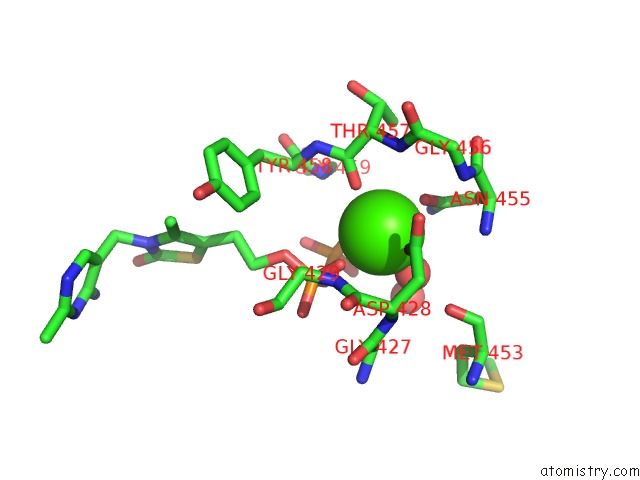
Mono view
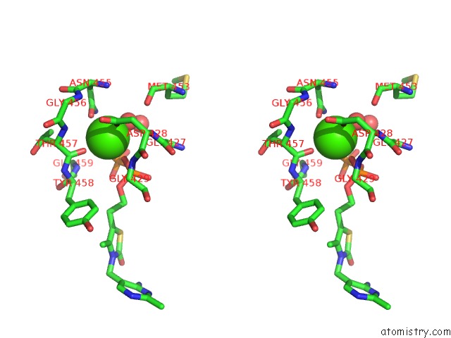
Stereo pair view
Mono view
Stereo pair view
A full contact list of Calcium with other atoms in the Ca binding
site number 1 of Crystal Structure of Benzoylformate Decarboxylase Mutant L403N within 5.0Å range:
|
Calcium binding site 2 out of 2 in 4gm0
Go back to
Calcium binding site 2 out
of 2 in the Crystal Structure of Benzoylformate Decarboxylase Mutant L403N
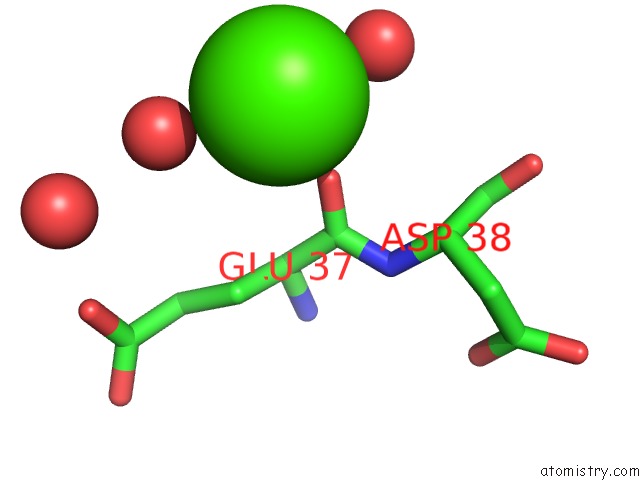
Mono view
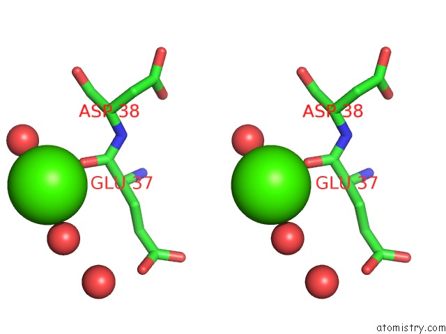
Stereo pair view
Mono view
Stereo pair view
A full contact list of Calcium with other atoms in the Ca binding
site number 2 of Crystal Structure of Benzoylformate Decarboxylase Mutant L403N within 5.0Å range:
|
Reference:
F.H.Andrews,
A.R.Tom,
P.R.Gunderman,
W.R.Novak,
M.J.Mcleish.
A Bulky Hydrophobic Residue Is Not Required to Maintain the V-Conformation of Enzyme-Bound Thiamin Diphosphate. Biochemistry V. 52 3028 2013.
ISSN: ISSN 0006-2960
PubMed: 23607689
DOI: 10.1021/BI400368J
Page generated: Sun Jul 14 07:15:44 2024
ISSN: ISSN 0006-2960
PubMed: 23607689
DOI: 10.1021/BI400368J
Last articles
Zn in 9J0NZn in 9J0O
Zn in 9J0P
Zn in 9FJX
Zn in 9EKB
Zn in 9C0F
Zn in 9CAH
Zn in 9CH0
Zn in 9CH3
Zn in 9CH1