Calcium »
PDB 4hvk-4i9x »
4i8h »
Calcium in PDB 4i8h: Bovine Trypsin at 0.75 Resolution
Enzymatic activity of Bovine Trypsin at 0.75 Resolution
All present enzymatic activity of Bovine Trypsin at 0.75 Resolution:
3.4.21.4;
3.4.21.4;
Protein crystallography data
The structure of Bovine Trypsin at 0.75 Resolution, PDB code: 4i8h
was solved by
Z.Dauter,
D.Liebschner,
M.Dauter,
A.Brzuszkiewicz,
with X-Ray Crystallography technique. A brief refinement statistics is given in the table below:
| Resolution Low / High (Å) | 30.00 / 0.75 |
| Space group | P 21 21 21 |
| Cell size a, b, c (Å), α, β, γ (°) | 54.342, 58.440, 66.476, 90.00, 90.00, 90.00 |
| R / Rfree (%) | n/a / 11.1 |
Calcium Binding Sites:
The binding sites of Calcium atom in the Bovine Trypsin at 0.75 Resolution
(pdb code 4i8h). This binding sites where shown within
5.0 Angstroms radius around Calcium atom.
In total only one binding site of Calcium was determined in the Bovine Trypsin at 0.75 Resolution, PDB code: 4i8h:
In total only one binding site of Calcium was determined in the Bovine Trypsin at 0.75 Resolution, PDB code: 4i8h:
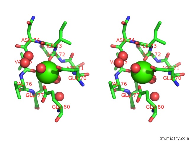
Calcium binding site 1 out of 1 in 4i8h
Go back to
Calcium binding site 1 out
of 1 in the Bovine Trypsin at 0.75 Resolution

Mono view
Stereo pair view
Mono view
Stereo pair view
A full contact list of Calcium with other atoms in the Ca binding
site number 1 of Bovine Trypsin at 0.75 Resolution within 5.0Å range:
|
Reference:
D.Liebschner,
M.Dauter,
A.Brzuszkiewicz,
Z.Dauter.
On the Reproducibility of Protein Crystal Structures: Five Atomic Resolution Structures of Trypsin. Acta Crystallogr.,Sect.D V. 69 1447 2013.
ISSN: ISSN 0907-4449
PubMed: 23897468
DOI: 10.1107/S0907444913009050
Page generated: Sun Jul 14 08:04:24 2024
ISSN: ISSN 0907-4449
PubMed: 23897468
DOI: 10.1107/S0907444913009050
Last articles
Zn in 9J0NZn in 9J0O
Zn in 9J0P
Zn in 9FJX
Zn in 9EKB
Zn in 9C0F
Zn in 9CAH
Zn in 9CH0
Zn in 9CH3
Zn in 9CH1