Calcium in PDB 4j3u: Crystal Structure of Barley Limit Dextrinase in Complex with Maltosyl- S-Betacyclodextrin
Enzymatic activity of Crystal Structure of Barley Limit Dextrinase in Complex with Maltosyl- S-Betacyclodextrin
All present enzymatic activity of Crystal Structure of Barley Limit Dextrinase in Complex with Maltosyl- S-Betacyclodextrin:
3.2.1.41;
Protein crystallography data
The structure of Crystal Structure of Barley Limit Dextrinase in Complex with Maltosyl- S-Betacyclodextrin, PDB code: 4j3u
was solved by
L.Sim,
M.S.Windahl,
M.S.Moeller,
A.Henriksen,
with X-Ray Crystallography technique. A brief refinement statistics is given in the table below:
Resolution Low / High (Å)
|
29.76 /
1.70
|
Space group
|
C 1 2 1
|
Cell size a, b, c (Å), α, β, γ (°)
|
195.430,
84.610,
121.790,
90.00,
119.91,
90.00
|
R / Rfree (%)
|
17.2 /
21.8
|
Other elements in 4j3u:
The structure of Crystal Structure of Barley Limit Dextrinase in Complex with Maltosyl- S-Betacyclodextrin also contains other interesting chemical elements:
Calcium Binding Sites:
The binding sites of Calcium atom in the Crystal Structure of Barley Limit Dextrinase in Complex with Maltosyl- S-Betacyclodextrin
(pdb code 4j3u). This binding sites where shown within
5.0 Angstroms radius around Calcium atom.
In total 4 binding sites of Calcium where determined in the
Crystal Structure of Barley Limit Dextrinase in Complex with Maltosyl- S-Betacyclodextrin, PDB code: 4j3u:
Jump to Calcium binding site number:
1;
2;
3;
4;
Calcium binding site 1 out
of 4 in 4j3u
Go back to
Calcium Binding Sites List in 4j3u
Calcium binding site 1 out
of 4 in the Crystal Structure of Barley Limit Dextrinase in Complex with Maltosyl- S-Betacyclodextrin
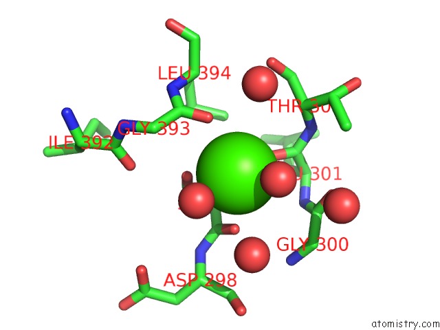
Mono view
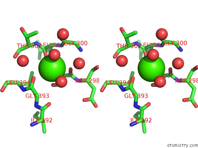
Stereo pair view
|
A full contact list of Calcium with other atoms in the Ca binding
site number 1 of Crystal Structure of Barley Limit Dextrinase in Complex with Maltosyl- S-Betacyclodextrin within 5.0Å range:
probe
|
atom
|
residue
|
distance (Å)
|
B
|
Occ
|
A:Ca930
b:11.2
occ:1.00
|
O
|
A:HOH1315
|
2.6
|
26.5
|
1.0
|
O
|
A:LEU301
|
2.7
|
10.3
|
1.0
|
O
|
A:HOH1361
|
2.7
|
24.2
|
1.0
|
O
|
A:GLY393
|
2.7
|
10.3
|
1.0
|
OG
|
A:SER297
|
2.8
|
9.7
|
1.0
|
O
|
A:SER297
|
2.8
|
8.8
|
1.0
|
C
|
A:SER297
|
3.6
|
9.5
|
1.0
|
C
|
A:GLY393
|
3.7
|
10.2
|
1.0
|
O
|
A:HOH1267
|
3.7
|
22.8
|
1.0
|
CA
|
A:SER297
|
3.7
|
9.2
|
1.0
|
CB
|
A:SER297
|
3.8
|
9.1
|
1.0
|
C
|
A:LEU301
|
3.8
|
9.0
|
1.0
|
O
|
A:HOH1112
|
4.1
|
12.0
|
1.0
|
CA
|
A:GLY393
|
4.1
|
10.1
|
1.0
|
C
|
A:GLY300
|
4.4
|
7.4
|
1.0
|
CA
|
A:THR302
|
4.4
|
9.4
|
1.0
|
O
|
A:GLY300
|
4.5
|
7.5
|
1.0
|
N
|
A:LEU301
|
4.6
|
7.6
|
1.0
|
CD2
|
A:LEU394
|
4.6
|
9.7
|
1.0
|
N
|
A:THR302
|
4.6
|
9.0
|
1.0
|
N
|
A:ASP298
|
4.7
|
10.3
|
1.0
|
N
|
A:LEU394
|
4.8
|
10.4
|
1.0
|
O
|
A:HOH1253
|
4.8
|
13.0
|
1.0
|
CA
|
A:LEU301
|
4.8
|
8.8
|
1.0
|
CA
|
A:GLY300
|
4.8
|
7.1
|
1.0
|
O
|
A:ILE392
|
4.9
|
12.1
|
1.0
|
|
Calcium binding site 2 out
of 4 in 4j3u
Go back to
Calcium Binding Sites List in 4j3u
Calcium binding site 2 out
of 4 in the Crystal Structure of Barley Limit Dextrinase in Complex with Maltosyl- S-Betacyclodextrin
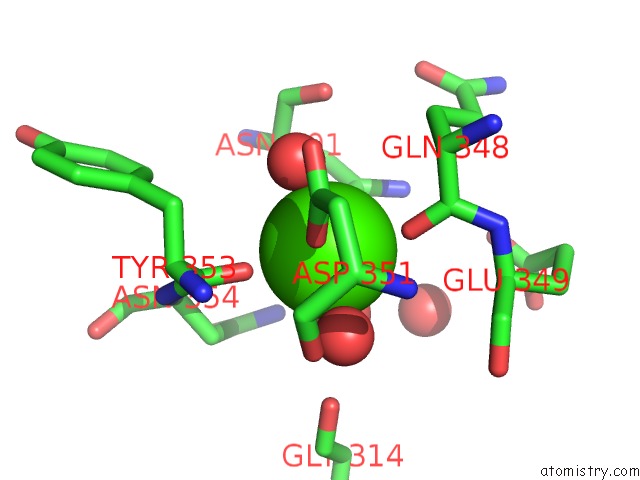
Mono view
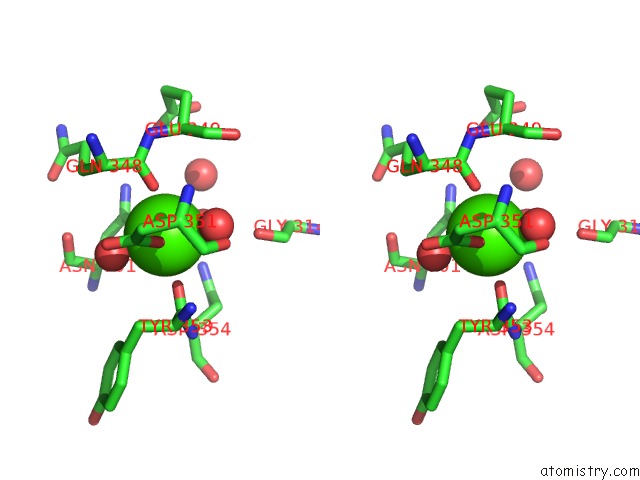
Stereo pair view
|
A full contact list of Calcium with other atoms in the Ca binding
site number 2 of Crystal Structure of Barley Limit Dextrinase in Complex with Maltosyl- S-Betacyclodextrin within 5.0Å range:
probe
|
atom
|
residue
|
distance (Å)
|
B
|
Occ
|
A:Ca931
b:12.6
occ:1.00
|
O
|
A:GLN348
|
2.2
|
16.9
|
1.0
|
O
|
A:TYR353
|
2.3
|
12.1
|
1.0
|
OD1
|
A:ASN701
|
2.4
|
11.4
|
1.0
|
O
|
A:HOH1002
|
2.5
|
14.0
|
1.0
|
OD1
|
A:ASP351
|
2.5
|
15.1
|
1.0
|
O
|
A:HOH1001
|
2.5
|
11.0
|
1.0
|
O
|
A:HOH1225
|
2.5
|
12.6
|
1.0
|
C
|
A:GLN348
|
3.4
|
17.8
|
1.0
|
C
|
A:TYR353
|
3.5
|
11.5
|
1.0
|
CG
|
A:ASP351
|
3.5
|
14.9
|
1.0
|
CG
|
A:ASN701
|
3.6
|
11.3
|
1.0
|
OD2
|
A:ASP351
|
3.8
|
15.8
|
1.0
|
CA
|
A:GLN348
|
4.1
|
17.9
|
1.0
|
CA
|
A:TYR353
|
4.2
|
12.3
|
1.0
|
O
|
A:GLY314
|
4.2
|
14.0
|
1.0
|
N
|
A:TYR353
|
4.2
|
13.2
|
1.0
|
ND2
|
A:ASN701
|
4.3
|
12.6
|
1.0
|
CB
|
A:TYR353
|
4.4
|
12.9
|
1.0
|
O
|
A:ASP351
|
4.4
|
13.9
|
1.0
|
N
|
A:GLU349
|
4.5
|
18.7
|
1.0
|
CB
|
A:GLN348
|
4.5
|
19.0
|
1.0
|
N
|
A:ASN354
|
4.5
|
10.8
|
1.0
|
ND2
|
A:ASN354
|
4.6
|
13.1
|
1.0
|
CA
|
A:ASN701
|
4.6
|
11.3
|
1.0
|
O
|
A:ASN701
|
4.6
|
12.1
|
1.0
|
O
|
A:HOH1153
|
4.6
|
21.0
|
1.0
|
CB
|
A:ASN701
|
4.7
|
11.2
|
1.0
|
CA
|
A:GLU349
|
4.7
|
19.2
|
1.0
|
CB
|
A:ASN354
|
4.7
|
11.8
|
1.0
|
N
|
A:ASP351
|
4.7
|
15.6
|
1.0
|
CB
|
A:ASP351
|
4.8
|
14.8
|
1.0
|
CA
|
A:ASN354
|
4.8
|
10.8
|
1.0
|
C
|
A:ASP351
|
4.8
|
14.4
|
1.0
|
C
|
A:GLU349
|
4.9
|
18.3
|
1.0
|
C
|
A:ASN701
|
4.9
|
12.0
|
1.0
|
|
Calcium binding site 3 out
of 4 in 4j3u
Go back to
Calcium Binding Sites List in 4j3u
Calcium binding site 3 out
of 4 in the Crystal Structure of Barley Limit Dextrinase in Complex with Maltosyl- S-Betacyclodextrin
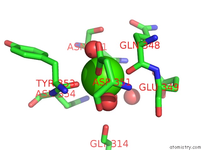
Mono view
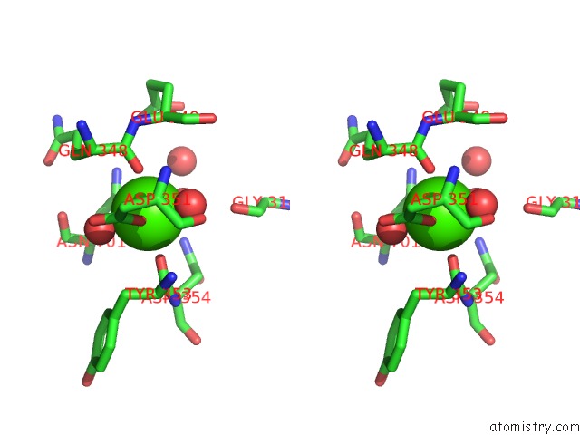
Stereo pair view
|
A full contact list of Calcium with other atoms in the Ca binding
site number 3 of Crystal Structure of Barley Limit Dextrinase in Complex with Maltosyl- S-Betacyclodextrin within 5.0Å range:
probe
|
atom
|
residue
|
distance (Å)
|
B
|
Occ
|
B:Ca922
b:12.3
occ:1.00
|
O
|
B:GLN348
|
2.2
|
16.9
|
1.0
|
O
|
B:TYR353
|
2.3
|
10.7
|
1.0
|
O
|
B:HOH1805
|
2.3
|
13.9
|
1.0
|
OD1
|
B:ASN701
|
2.4
|
12.9
|
1.0
|
O
|
B:HOH1784
|
2.4
|
13.5
|
1.0
|
O
|
B:HOH1206
|
2.5
|
13.9
|
1.0
|
OD1
|
B:ASP351
|
2.5
|
14.0
|
1.0
|
C
|
B:GLN348
|
3.4
|
17.4
|
1.0
|
C
|
B:TYR353
|
3.4
|
9.6
|
1.0
|
CG
|
B:ASP351
|
3.5
|
13.4
|
1.0
|
CG
|
B:ASN701
|
3.6
|
13.3
|
1.0
|
OD2
|
B:ASP351
|
3.8
|
14.8
|
1.0
|
CA
|
B:GLN348
|
4.1
|
17.4
|
1.0
|
CA
|
B:TYR353
|
4.1
|
10.0
|
1.0
|
N
|
B:TYR353
|
4.2
|
10.9
|
1.0
|
ND2
|
B:ASN701
|
4.3
|
14.9
|
1.0
|
O
|
B:GLY314
|
4.3
|
13.7
|
1.0
|
CB
|
B:TYR353
|
4.3
|
9.6
|
1.0
|
CB
|
B:GLN348
|
4.5
|
17.7
|
1.0
|
N
|
B:GLU349
|
4.5
|
17.3
|
1.0
|
N
|
B:ASN354
|
4.5
|
10.3
|
1.0
|
O
|
B:ASP351
|
4.5
|
12.4
|
1.0
|
CA
|
B:ASN701
|
4.6
|
13.5
|
1.0
|
O
|
B:ASN701
|
4.6
|
12.7
|
1.0
|
ND2
|
B:ASN354
|
4.6
|
11.5
|
1.0
|
O
|
B:HOH1060
|
4.7
|
22.1
|
1.0
|
CB
|
B:ASN354
|
4.7
|
10.8
|
1.0
|
CB
|
B:ASN701
|
4.7
|
13.6
|
1.0
|
CA
|
B:GLU349
|
4.7
|
18.1
|
1.0
|
N
|
B:ASP351
|
4.8
|
14.0
|
1.0
|
CA
|
B:ASN354
|
4.8
|
10.0
|
1.0
|
CB
|
B:ASP351
|
4.8
|
13.3
|
1.0
|
C
|
B:ASP351
|
4.9
|
12.9
|
1.0
|
C
|
B:ASN701
|
4.9
|
12.9
|
1.0
|
|
Calcium binding site 4 out
of 4 in 4j3u
Go back to
Calcium Binding Sites List in 4j3u
Calcium binding site 4 out
of 4 in the Crystal Structure of Barley Limit Dextrinase in Complex with Maltosyl- S-Betacyclodextrin
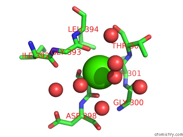
Mono view
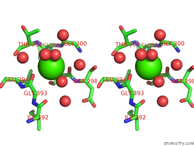
Stereo pair view
|
A full contact list of Calcium with other atoms in the Ca binding
site number 4 of Crystal Structure of Barley Limit Dextrinase in Complex with Maltosyl- S-Betacyclodextrin within 5.0Å range:
probe
|
atom
|
residue
|
distance (Å)
|
B
|
Occ
|
B:Ca923
b:10.9
occ:1.00
|
O
|
B:HOH1326
|
2.5
|
29.6
|
1.0
|
O
|
B:LEU301
|
2.6
|
9.5
|
1.0
|
OG
|
B:SER297
|
2.7
|
9.7
|
1.0
|
O
|
B:GLY393
|
2.7
|
10.7
|
1.0
|
O
|
B:HOH1145
|
2.8
|
15.8
|
1.0
|
O
|
B:SER297
|
2.9
|
8.5
|
1.0
|
C
|
B:SER297
|
3.6
|
10.1
|
1.0
|
C
|
B:GLY393
|
3.7
|
10.2
|
1.0
|
C
|
B:LEU301
|
3.8
|
8.4
|
1.0
|
CA
|
B:SER297
|
3.8
|
9.8
|
1.0
|
CB
|
B:SER297
|
3.8
|
9.9
|
1.0
|
O
|
B:HOH1244
|
3.9
|
16.0
|
1.0
|
O
|
B:HOH1097
|
4.1
|
13.9
|
1.0
|
CA
|
B:GLY393
|
4.1
|
10.7
|
1.0
|
C
|
B:GLY300
|
4.4
|
8.6
|
1.0
|
CA
|
B:THR302
|
4.4
|
8.3
|
1.0
|
O
|
B:GLY300
|
4.4
|
7.7
|
1.0
|
O
|
B:HOH1187
|
4.5
|
12.5
|
1.0
|
N
|
B:THR302
|
4.5
|
7.7
|
1.0
|
CD2
|
B:LEU394
|
4.6
|
8.6
|
1.0
|
N
|
B:LEU301
|
4.6
|
8.5
|
1.0
|
N
|
B:ASP298
|
4.7
|
10.4
|
1.0
|
N
|
B:LEU394
|
4.8
|
9.8
|
1.0
|
CA
|
B:LEU301
|
4.8
|
8.6
|
1.0
|
CA
|
B:GLY300
|
4.8
|
9.7
|
1.0
|
O
|
B:HOH1511
|
4.8
|
27.6
|
1.0
|
O
|
B:ILE392
|
4.9
|
12.6
|
1.0
|
O
|
B:HOH1533
|
5.0
|
22.6
|
1.0
|
|
Reference:
L.Sim,
M.S.Windahl,
M.S.Moeller,
A.Henriksen.
Crystal Structure of Barley Limit Dextrinase in Complex with Maltosyl-S-Betacyclodextrin To Be Published.
Page generated: Sun Jul 14 08:24:54 2024
|