Calcium »
PDB 4jo1-4k0g »
4jso »
Calcium in PDB 4jso: The X-Ray Crystal Structure of A Thermophilic Cellobiose Binding Protein Bound with Laminaripentaose
Protein crystallography data
The structure of The X-Ray Crystal Structure of A Thermophilic Cellobiose Binding Protein Bound with Laminaripentaose, PDB code: 4jso
was solved by
P.Munshi,
M.J.Cuneo,
with X-Ray Crystallography technique. A brief refinement statistics is given in the table below:
| Resolution Low / High (Å) | 48.08 / 2.07 |
| Space group | P 21 21 21 |
| Cell size a, b, c (Å), α, β, γ (°) | 56.922, 89.785, 108.321, 90.00, 90.00, 90.00 |
| R / Rfree (%) | 18.7 / 22.1 |
Calcium Binding Sites:
The binding sites of Calcium atom in the The X-Ray Crystal Structure of A Thermophilic Cellobiose Binding Protein Bound with Laminaripentaose
(pdb code 4jso). This binding sites where shown within
5.0 Angstroms radius around Calcium atom.
In total only one binding site of Calcium was determined in the The X-Ray Crystal Structure of A Thermophilic Cellobiose Binding Protein Bound with Laminaripentaose, PDB code: 4jso:
In total only one binding site of Calcium was determined in the The X-Ray Crystal Structure of A Thermophilic Cellobiose Binding Protein Bound with Laminaripentaose, PDB code: 4jso:
Calcium binding site 1 out of 1 in 4jso
Go back to
Calcium binding site 1 out
of 1 in the The X-Ray Crystal Structure of A Thermophilic Cellobiose Binding Protein Bound with Laminaripentaose
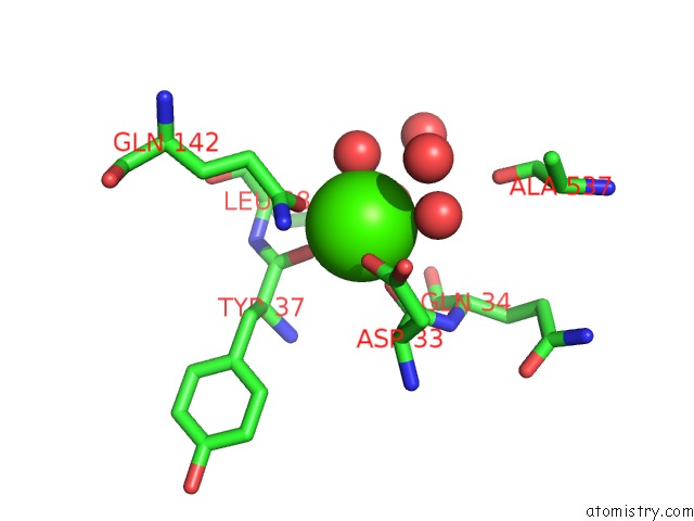
Mono view
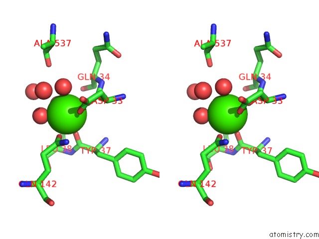
Stereo pair view
Mono view
Stereo pair view
A full contact list of Calcium with other atoms in the Ca binding
site number 1 of The X-Ray Crystal Structure of A Thermophilic Cellobiose Binding Protein Bound with Laminaripentaose within 5.0Å range:
|
Reference:
P.Munshi,
C.B.Stanley,
S.Ghimire-Rijal,
X.Lu,
D.A.Myles,
M.J.Cuneo.
Molecular Details of Ligand Selectivity Determinants in A Promiscuous Beta-Glucan Periplasmic Binding Protein. Bmc Struct.Biol. V. 13 18 2013.
ISSN: ESSN 1472-6807
PubMed: 24090243
DOI: 10.1186/1472-6807-13-18
Page generated: Sun Jul 14 08:38:20 2024
ISSN: ESSN 1472-6807
PubMed: 24090243
DOI: 10.1186/1472-6807-13-18
Last articles
Zn in 9J0NZn in 9J0O
Zn in 9J0P
Zn in 9FJX
Zn in 9EKB
Zn in 9C0F
Zn in 9CAH
Zn in 9CH0
Zn in 9CH3
Zn in 9CH1