Calcium »
PDB 4jo1-4k0g »
4jwp »
Calcium in PDB 4jwp: Crystal Structure of Ribosomal-Protein-Alanine N-Acetyltransferase From Brucella Melitensis in Complex with Acetyl Coa
Protein crystallography data
The structure of Crystal Structure of Ribosomal-Protein-Alanine N-Acetyltransferase From Brucella Melitensis in Complex with Acetyl Coa, PDB code: 4jwp
was solved by
Ssgcid,
Seattle Structural Genomics Center For Infectious Disease(Ssgcid),
with X-Ray Crystallography technique. A brief refinement statistics is given in the table below:
| Resolution Low / High (Å) | 41.58 / 2.00 |
| Space group | C 1 2 1 |
| Cell size a, b, c (Å), α, β, γ (°) | 72.420, 74.380, 67.550, 90.00, 91.62, 90.00 |
| R / Rfree (%) | 17.3 / 22.5 |
Other elements in 4jwp:
The structure of Crystal Structure of Ribosomal-Protein-Alanine N-Acetyltransferase From Brucella Melitensis in Complex with Acetyl Coa also contains other interesting chemical elements:
| Chlorine | (Cl) | 1 atom |
Calcium Binding Sites:
The binding sites of Calcium atom in the Crystal Structure of Ribosomal-Protein-Alanine N-Acetyltransferase From Brucella Melitensis in Complex with Acetyl Coa
(pdb code 4jwp). This binding sites where shown within
5.0 Angstroms radius around Calcium atom.
In total 4 binding sites of Calcium where determined in the Crystal Structure of Ribosomal-Protein-Alanine N-Acetyltransferase From Brucella Melitensis in Complex with Acetyl Coa, PDB code: 4jwp:
Jump to Calcium binding site number: 1; 2; 3; 4;
In total 4 binding sites of Calcium where determined in the Crystal Structure of Ribosomal-Protein-Alanine N-Acetyltransferase From Brucella Melitensis in Complex with Acetyl Coa, PDB code: 4jwp:
Jump to Calcium binding site number: 1; 2; 3; 4;
Calcium binding site 1 out of 4 in 4jwp
Go back to
Calcium binding site 1 out
of 4 in the Crystal Structure of Ribosomal-Protein-Alanine N-Acetyltransferase From Brucella Melitensis in Complex with Acetyl Coa
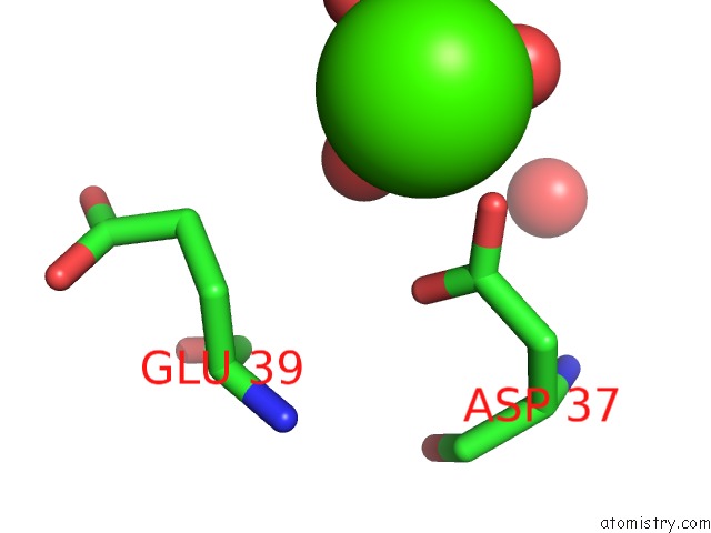
Mono view

Stereo pair view
Mono view
Stereo pair view
A full contact list of Calcium with other atoms in the Ca binding
site number 1 of Crystal Structure of Ribosomal-Protein-Alanine N-Acetyltransferase From Brucella Melitensis in Complex with Acetyl Coa within 5.0Å range:
|
Calcium binding site 2 out of 4 in 4jwp
Go back to
Calcium binding site 2 out
of 4 in the Crystal Structure of Ribosomal-Protein-Alanine N-Acetyltransferase From Brucella Melitensis in Complex with Acetyl Coa

Mono view
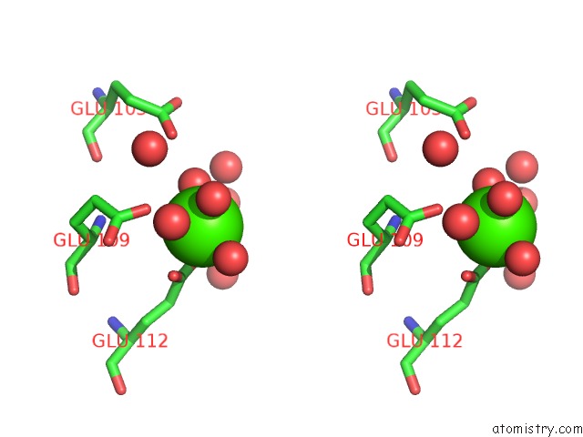
Stereo pair view
Mono view
Stereo pair view
A full contact list of Calcium with other atoms in the Ca binding
site number 2 of Crystal Structure of Ribosomal-Protein-Alanine N-Acetyltransferase From Brucella Melitensis in Complex with Acetyl Coa within 5.0Å range:
|
Calcium binding site 3 out of 4 in 4jwp
Go back to
Calcium binding site 3 out
of 4 in the Crystal Structure of Ribosomal-Protein-Alanine N-Acetyltransferase From Brucella Melitensis in Complex with Acetyl Coa
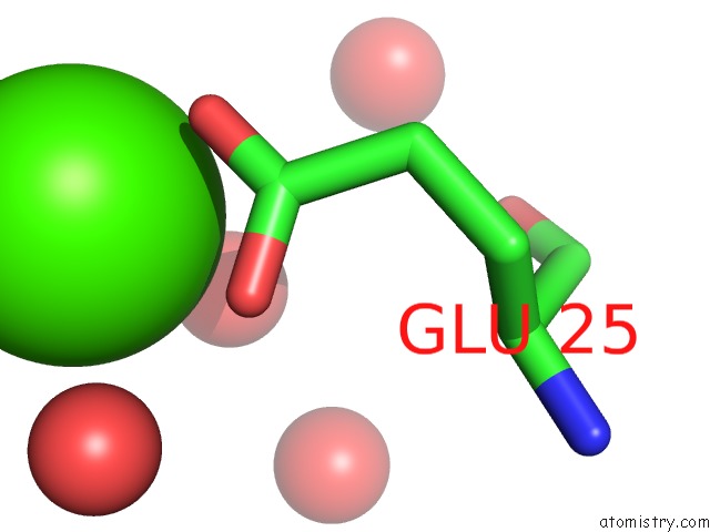
Mono view
Stereo pair view
Mono view
Stereo pair view
A full contact list of Calcium with other atoms in the Ca binding
site number 3 of Crystal Structure of Ribosomal-Protein-Alanine N-Acetyltransferase From Brucella Melitensis in Complex with Acetyl Coa within 5.0Å range:
|
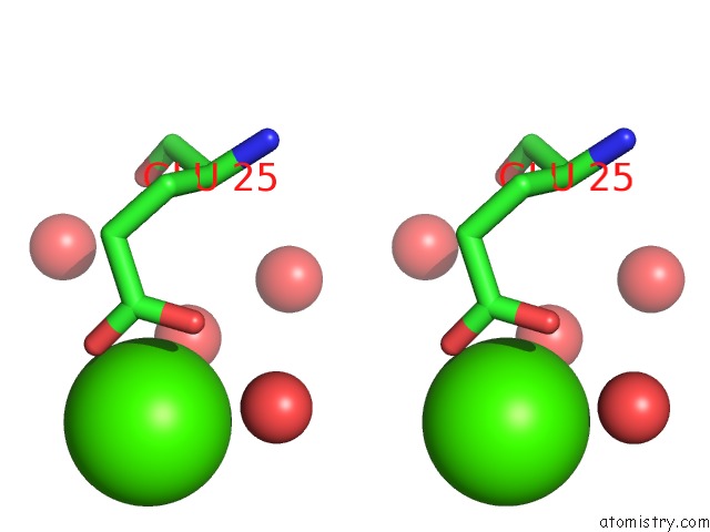
Calcium binding site 4 out of 4 in 4jwp
Go back to
Calcium binding site 4 out
of 4 in the Crystal Structure of Ribosomal-Protein-Alanine N-Acetyltransferase From Brucella Melitensis in Complex with Acetyl Coa

Mono view

Stereo pair view
Mono view
Stereo pair view
A full contact list of Calcium with other atoms in the Ca binding
site number 4 of Crystal Structure of Ribosomal-Protein-Alanine N-Acetyltransferase From Brucella Melitensis in Complex with Acetyl Coa within 5.0Å range:
|
Reference:
Seattle Structural Genomics Center For Infectious Disease(Ssgcid),
J.Abendroth,
T.Arakaki,
D.Lorimer,
T.E.Edwards.
Crystal Structure of Ribosomal-Protein-Alanine N-Acetyltransferase From Brucella Melitensis in Complex with Acetyl Coa To Be Published.
Page generated: Tue Jul 8 23:15:54 2025
Last articles
Cl in 8B7WCl in 8B56
Cl in 8B7B
Cl in 8B7A
Cl in 8B78
Cl in 8B6T
Cl in 8B6S
Cl in 8B75
Cl in 8B6P
Cl in 8B6R