Calcium »
PDB 4jo1-4k0g »
4jyo »
Calcium in PDB 4jyo: Structural Basis For Angiopoietin-1 Mediated Signaling Initiation
Protein crystallography data
The structure of Structural Basis For Angiopoietin-1 Mediated Signaling Initiation, PDB code: 4jyo
was solved by
X.Yu,
T.C.M.Seegar,
A.C.Dalton,
D.Tzvetkova-Robev,
Y.Goldgur,
D.B.Nikolov,
W.A.Barton,
with X-Ray Crystallography technique. A brief refinement statistics is given in the table below:
| Resolution Low / High (Å) | 30.00 / 2.50 |
| Space group | P 64 2 2 |
| Cell size a, b, c (Å), α, β, γ (°) | 80.968, 80.968, 187.216, 90.00, 90.00, 120.00 |
| R / Rfree (%) | 18.6 / 24.2 |
Calcium Binding Sites:
The binding sites of Calcium atom in the Structural Basis For Angiopoietin-1 Mediated Signaling Initiation
(pdb code 4jyo). This binding sites where shown within
5.0 Angstroms radius around Calcium atom.
In total only one binding site of Calcium was determined in the Structural Basis For Angiopoietin-1 Mediated Signaling Initiation, PDB code: 4jyo:
In total only one binding site of Calcium was determined in the Structural Basis For Angiopoietin-1 Mediated Signaling Initiation, PDB code: 4jyo:
Calcium binding site 1 out of 1 in 4jyo
Go back to
Calcium binding site 1 out
of 1 in the Structural Basis For Angiopoietin-1 Mediated Signaling Initiation
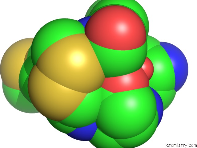
Mono view
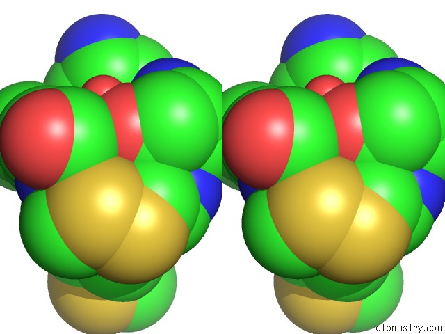
Stereo pair view
Mono view
Stereo pair view
A full contact list of Calcium with other atoms in the Ca binding
site number 1 of Structural Basis For Angiopoietin-1 Mediated Signaling Initiation within 5.0Å range:
|
Reference:
X.Yu,
T.C.Seegar,
A.C.Dalton,
D.Tzvetkova-Robev,
Y.Goldgur,
K.R.Rajashankar,
D.B.Nikolov,
W.A.Barton.
Structural Basis For Angiopoietin-1-Mediated Signaling Initiation. Proc.Natl.Acad.Sci.Usa V. 110 7205 2013.
ISSN: ISSN 0027-8424
PubMed: 23592718
DOI: 10.1073/PNAS.1216890110
Page generated: Tue Jul 8 23:16:40 2025
ISSN: ISSN 0027-8424
PubMed: 23592718
DOI: 10.1073/PNAS.1216890110
Last articles
Cl in 5GTHCl in 5GTD
Cl in 5GT3
Cl in 5GT0
Cl in 5GTC
Cl in 5GRQ
Cl in 5GRN
Cl in 5GSU
Cl in 5GS8
Cl in 5GQQ