Calcium »
PDB 4k9m-4ko1 »
4kif »
Calcium in PDB 4kif: Crystal Structure of Methyltransferase From Streptomyces Hygroscopicus Complexed with Phenylpyruvic Acid
Protein crystallography data
The structure of Crystal Structure of Methyltransferase From Streptomyces Hygroscopicus Complexed with Phenylpyruvic Acid, PDB code: 4kif
was solved by
Y.C.Liu,
X.W.Zou,
H.C.Chan,
C.J.Huang,
T.L.Li,
with X-Ray Crystallography technique. A brief refinement statistics is given in the table below:
| Resolution Low / High (Å) | 29.43 / 2.50 |
| Space group | P 21 21 21 |
| Cell size a, b, c (Å), α, β, γ (°) | 57.431, 88.708, 137.005, 90.00, 90.00, 90.00 |
| R / Rfree (%) | 17 / 24.8 |
Other elements in 4kif:
The structure of Crystal Structure of Methyltransferase From Streptomyces Hygroscopicus Complexed with Phenylpyruvic Acid also contains other interesting chemical elements:
| Iron | (Fe) | 2 atoms |
Calcium Binding Sites:
The binding sites of Calcium atom in the Crystal Structure of Methyltransferase From Streptomyces Hygroscopicus Complexed with Phenylpyruvic Acid
(pdb code 4kif). This binding sites where shown within
5.0 Angstroms radius around Calcium atom.
In total 4 binding sites of Calcium where determined in the Crystal Structure of Methyltransferase From Streptomyces Hygroscopicus Complexed with Phenylpyruvic Acid, PDB code: 4kif:
Jump to Calcium binding site number: 1; 2; 3; 4;
In total 4 binding sites of Calcium where determined in the Crystal Structure of Methyltransferase From Streptomyces Hygroscopicus Complexed with Phenylpyruvic Acid, PDB code: 4kif:
Jump to Calcium binding site number: 1; 2; 3; 4;
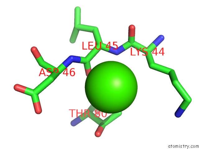
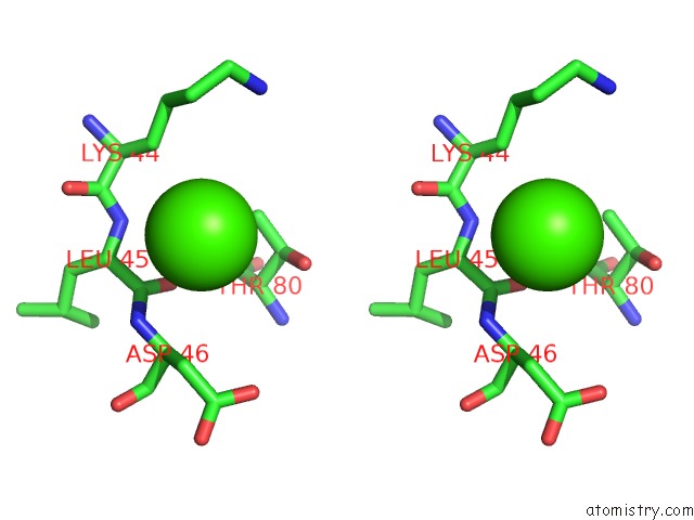
Calcium binding site 1 out of 4 in 4kif
Go back to
Calcium binding site 1 out
of 4 in the Crystal Structure of Methyltransferase From Streptomyces Hygroscopicus Complexed with Phenylpyruvic Acid
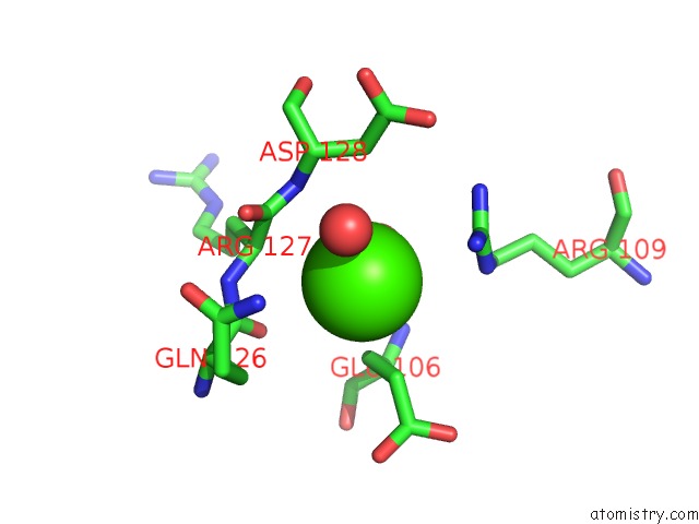
Mono view
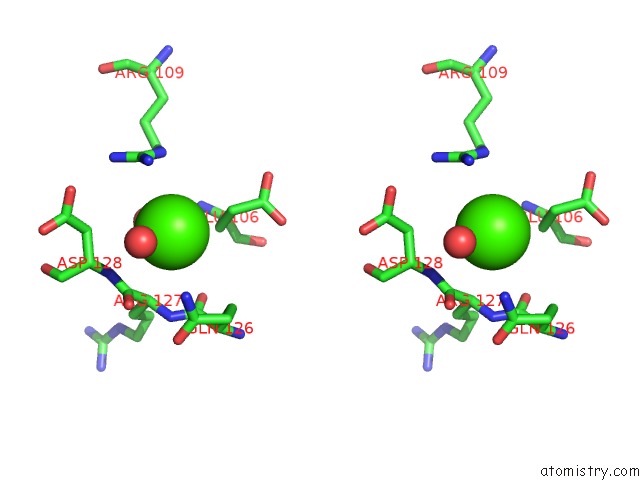
Stereo pair view
Mono view
Stereo pair view
A full contact list of Calcium with other atoms in the Ca binding
site number 1 of Crystal Structure of Methyltransferase From Streptomyces Hygroscopicus Complexed with Phenylpyruvic Acid within 5.0Å range:
|
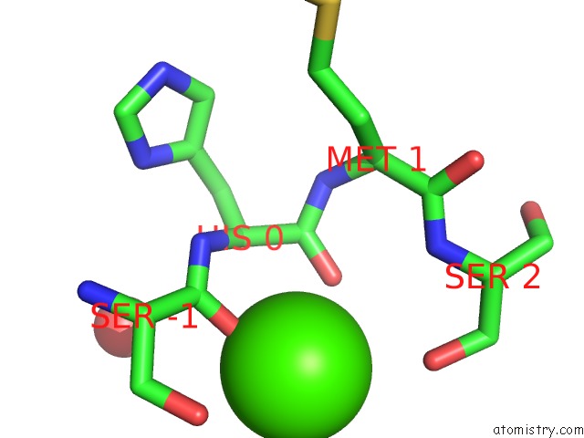
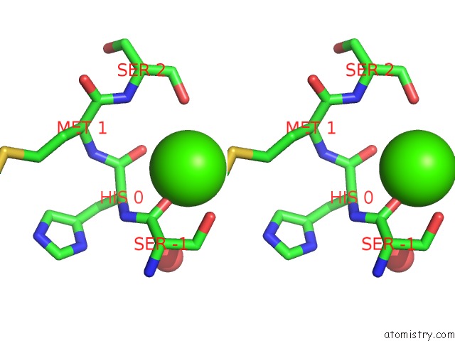
Calcium binding site 2 out of 4 in 4kif
Go back to
Calcium binding site 2 out
of 4 in the Crystal Structure of Methyltransferase From Streptomyces Hygroscopicus Complexed with Phenylpyruvic Acid
Mono view
Stereo pair view
Mono view
Stereo pair view
A full contact list of Calcium with other atoms in the Ca binding
site number 2 of Crystal Structure of Methyltransferase From Streptomyces Hygroscopicus Complexed with Phenylpyruvic Acid within 5.0Å range:
|
Calcium binding site 3 out of 4 in 4kif
Go back to
Calcium binding site 3 out
of 4 in the Crystal Structure of Methyltransferase From Streptomyces Hygroscopicus Complexed with Phenylpyruvic Acid
Mono view
Stereo pair view
Mono view
Stereo pair view
A full contact list of Calcium with other atoms in the Ca binding
site number 3 of Crystal Structure of Methyltransferase From Streptomyces Hygroscopicus Complexed with Phenylpyruvic Acid within 5.0Å range:
|
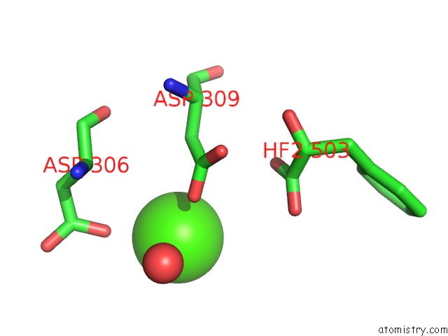
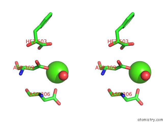
Calcium binding site 4 out of 4 in 4kif
Go back to
Calcium binding site 4 out
of 4 in the Crystal Structure of Methyltransferase From Streptomyces Hygroscopicus Complexed with Phenylpyruvic Acid
Mono view
Stereo pair view
Mono view
Stereo pair view
A full contact list of Calcium with other atoms in the Ca binding
site number 4 of Crystal Structure of Methyltransferase From Streptomyces Hygroscopicus Complexed with Phenylpyruvic Acid within 5.0Å range:
|
Reference:
X.W.Zou,
Y.C.Liu,
N.S.Hsu,
C.J.Huang,
S.Y.Lyu,
H.C.Chan,
C.Y.Chang,
H.W.Yeh,
K.H.Lin,
C.J.Wu,
M.D.Tsai,
T.L.Li.
Structure and Mechanism of A Nonhaem-Iron Sam-Dependent C-Methyltransferase and Its Engineering to A Hydratase and An O-Methyltransferase Acta Crystallogr.,Sect.D V. 70 1549 2014.
ISSN: ISSN 0907-4449
PubMed: 24914966
DOI: 10.1107/S1399004714005239
Page generated: Sun Jul 14 09:07:01 2024
ISSN: ISSN 0907-4449
PubMed: 24914966
DOI: 10.1107/S1399004714005239
Last articles
Zn in 9J0NZn in 9J0O
Zn in 9J0P
Zn in 9FJX
Zn in 9EKB
Zn in 9C0F
Zn in 9CAH
Zn in 9CH0
Zn in 9CH3
Zn in 9CH1