Calcium »
PDB 4ko2-4l19 »
4ktr »
Calcium in PDB 4ktr: Crystal Structure of 2-O-Alpha-Glucosylglycerol Phosphorylase in Complex with Isofagomine and Glycerol
Protein crystallography data
The structure of Crystal Structure of 2-O-Alpha-Glucosylglycerol Phosphorylase in Complex with Isofagomine and Glycerol, PDB code: 4ktr
was solved by
K.K.Touhara,
T.Nihira,
M.Kitaoka,
H.Nakai,
S.Fushinobu,
with X-Ray Crystallography technique. A brief refinement statistics is given in the table below:
| Resolution Low / High (Å) | 48.99 / 2.30 |
| Space group | P 1 21 1 |
| Cell size a, b, c (Å), α, β, γ (°) | 109.003, 263.213, 138.793, 90.00, 105.45, 90.00 |
| R / Rfree (%) | 16.7 / 22.8 |
Calcium Binding Sites:
The binding sites of Calcium atom in the Crystal Structure of 2-O-Alpha-Glucosylglycerol Phosphorylase in Complex with Isofagomine and Glycerol
(pdb code 4ktr). This binding sites where shown within
5.0 Angstroms radius around Calcium atom.
In total 7 binding sites of Calcium where determined in the Crystal Structure of 2-O-Alpha-Glucosylglycerol Phosphorylase in Complex with Isofagomine and Glycerol, PDB code: 4ktr:
Jump to Calcium binding site number: 1; 2; 3; 4; 5; 6; 7;
In total 7 binding sites of Calcium where determined in the Crystal Structure of 2-O-Alpha-Glucosylglycerol Phosphorylase in Complex with Isofagomine and Glycerol, PDB code: 4ktr:
Jump to Calcium binding site number: 1; 2; 3; 4; 5; 6; 7;
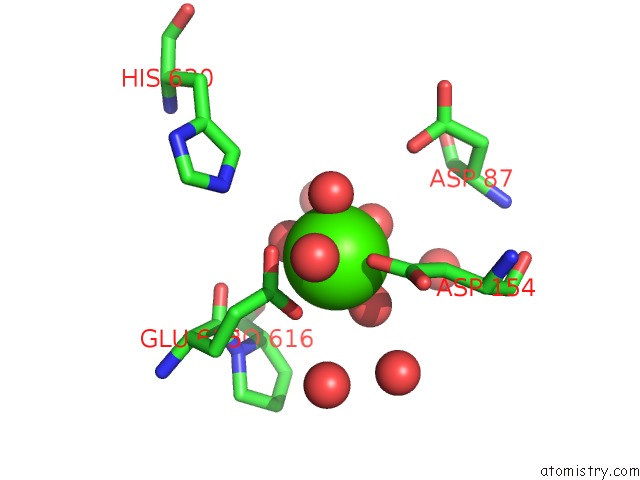
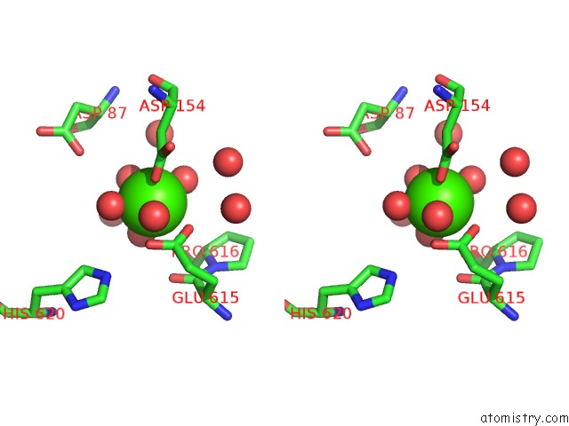
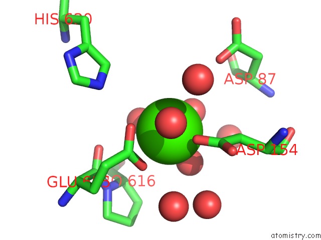
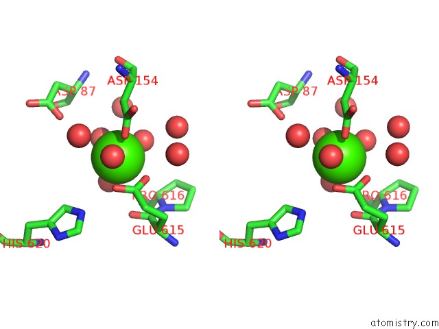
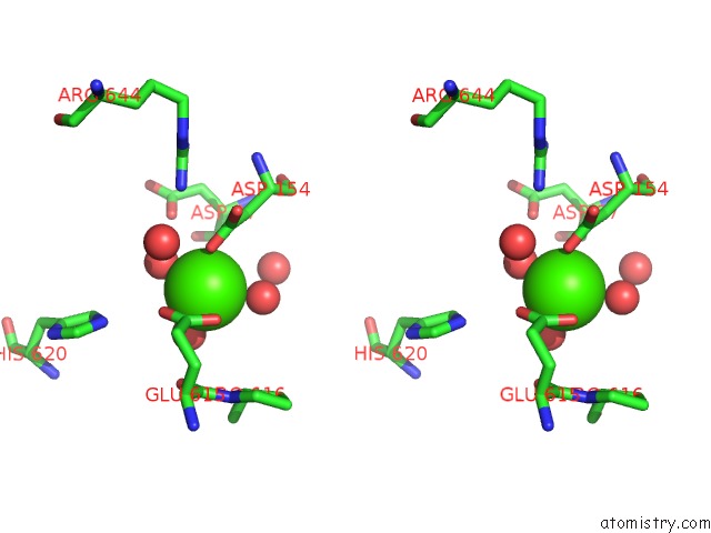
Calcium binding site 1 out of 7 in 4ktr
Go back to
Calcium binding site 1 out
of 7 in the Crystal Structure of 2-O-Alpha-Glucosylglycerol Phosphorylase in Complex with Isofagomine and Glycerol
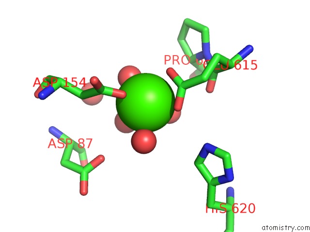
Mono view
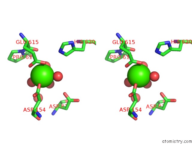
Stereo pair view
Mono view
Stereo pair view
A full contact list of Calcium with other atoms in the Ca binding
site number 1 of Crystal Structure of 2-O-Alpha-Glucosylglycerol Phosphorylase in Complex with Isofagomine and Glycerol within 5.0Å range:
|
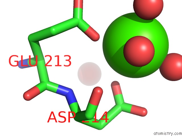
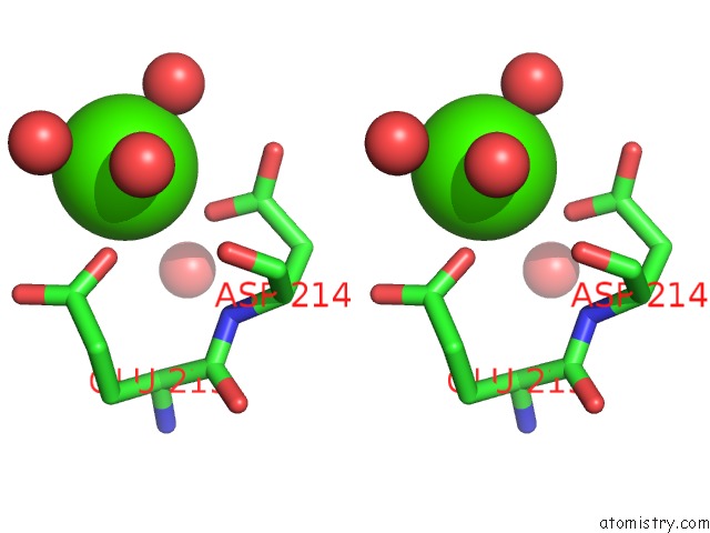
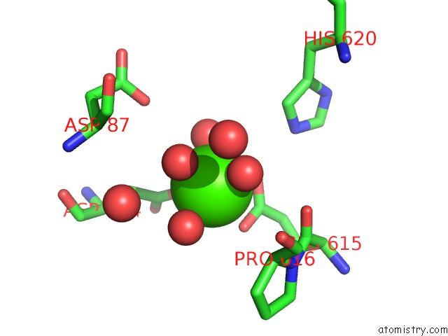
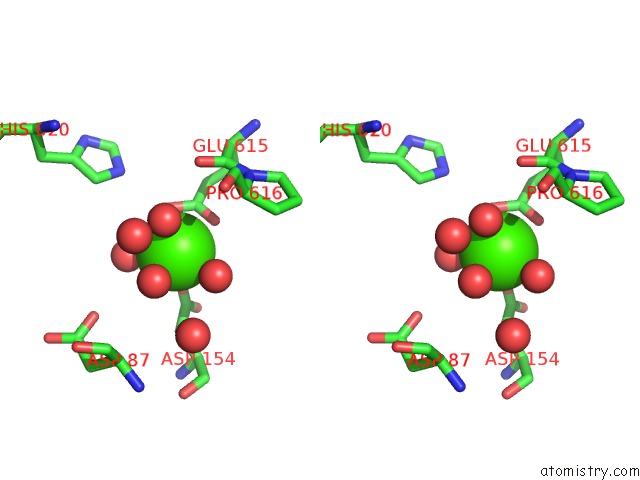
Calcium binding site 2 out of 7 in 4ktr
Go back to
Calcium binding site 2 out
of 7 in the Crystal Structure of 2-O-Alpha-Glucosylglycerol Phosphorylase in Complex with Isofagomine and Glycerol
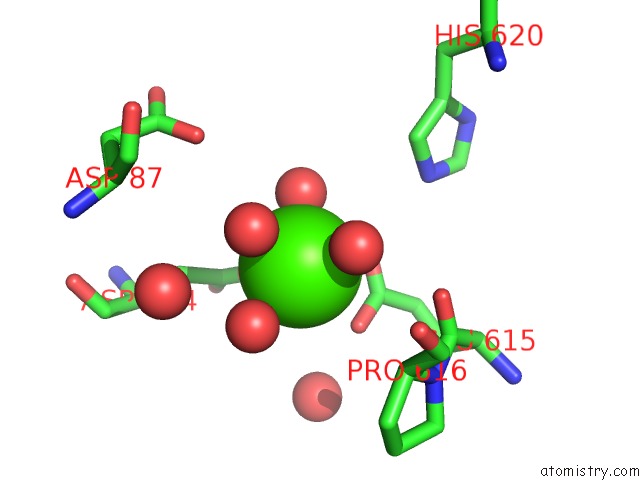
Mono view
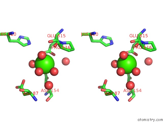
Stereo pair view
Mono view
Stereo pair view
A full contact list of Calcium with other atoms in the Ca binding
site number 2 of Crystal Structure of 2-O-Alpha-Glucosylglycerol Phosphorylase in Complex with Isofagomine and Glycerol within 5.0Å range:
|
Calcium binding site 3 out of 7 in 4ktr
Go back to
Calcium binding site 3 out
of 7 in the Crystal Structure of 2-O-Alpha-Glucosylglycerol Phosphorylase in Complex with Isofagomine and Glycerol
Mono view
Stereo pair view
Mono view
Stereo pair view
A full contact list of Calcium with other atoms in the Ca binding
site number 3 of Crystal Structure of 2-O-Alpha-Glucosylglycerol Phosphorylase in Complex with Isofagomine and Glycerol within 5.0Å range:
|
Calcium binding site 4 out of 7 in 4ktr
Go back to
Calcium binding site 4 out
of 7 in the Crystal Structure of 2-O-Alpha-Glucosylglycerol Phosphorylase in Complex with Isofagomine and Glycerol
Mono view
Stereo pair view
Mono view
Stereo pair view
A full contact list of Calcium with other atoms in the Ca binding
site number 4 of Crystal Structure of 2-O-Alpha-Glucosylglycerol Phosphorylase in Complex with Isofagomine and Glycerol within 5.0Å range:
|
Calcium binding site 5 out of 7 in 4ktr
Go back to
Calcium binding site 5 out
of 7 in the Crystal Structure of 2-O-Alpha-Glucosylglycerol Phosphorylase in Complex with Isofagomine and Glycerol
Mono view
Stereo pair view
Mono view
Stereo pair view
A full contact list of Calcium with other atoms in the Ca binding
site number 5 of Crystal Structure of 2-O-Alpha-Glucosylglycerol Phosphorylase in Complex with Isofagomine and Glycerol within 5.0Å range:
|
Calcium binding site 6 out of 7 in 4ktr
Go back to
Calcium binding site 6 out
of 7 in the Crystal Structure of 2-O-Alpha-Glucosylglycerol Phosphorylase in Complex with Isofagomine and Glycerol
Mono view
Stereo pair view
Mono view
Stereo pair view
A full contact list of Calcium with other atoms in the Ca binding
site number 6 of Crystal Structure of 2-O-Alpha-Glucosylglycerol Phosphorylase in Complex with Isofagomine and Glycerol within 5.0Å range:
|
Calcium binding site 7 out of 7 in 4ktr
Go back to
Calcium binding site 7 out
of 7 in the Crystal Structure of 2-O-Alpha-Glucosylglycerol Phosphorylase in Complex with Isofagomine and Glycerol

Mono view
Stereo pair view
Mono view
Stereo pair view
A full contact list of Calcium with other atoms in the Ca binding
site number 7 of Crystal Structure of 2-O-Alpha-Glucosylglycerol Phosphorylase in Complex with Isofagomine and Glycerol within 5.0Å range:
|
Reference:
K.K.Touhara,
T.Nihira,
M.Kitaoka,
H.Nakai,
S.Fushinobu.
Structural Basis For Reversible Phosphorolysis and Hydrolysis Reactions of 2-O-Alpha-Glucosylglycerol Phosphorylase J.Biol.Chem. 2014.
ISSN: ESSN 1083-351X
PubMed: 24828502
DOI: 10.1074/JBC.M114.573212
Page generated: Sun Jul 14 09:17:43 2024
ISSN: ESSN 1083-351X
PubMed: 24828502
DOI: 10.1074/JBC.M114.573212
Last articles
Zn in 9J0NZn in 9J0O
Zn in 9J0P
Zn in 9FJX
Zn in 9EKB
Zn in 9C0F
Zn in 9CAH
Zn in 9CH0
Zn in 9CH3
Zn in 9CH1