Calcium »
PDB 4ko2-4l19 »
4kxa »
Calcium in PDB 4kxa: Crystal Structure of Human Aminopeptidase A Complexed with Aspartate and Calcium
Enzymatic activity of Crystal Structure of Human Aminopeptidase A Complexed with Aspartate and Calcium
All present enzymatic activity of Crystal Structure of Human Aminopeptidase A Complexed with Aspartate and Calcium:
3.4.11.7;
3.4.11.7;
Protein crystallography data
The structure of Crystal Structure of Human Aminopeptidase A Complexed with Aspartate and Calcium, PDB code: 4kxa
was solved by
Y.Yang,
C.Liu,
Y.Y.Lin,
F.Li,
with X-Ray Crystallography technique. A brief refinement statistics is given in the table below:
| Resolution Low / High (Å) | 48.57 / 2.40 |
| Space group | P 64 2 2 |
| Cell size a, b, c (Å), α, β, γ (°) | 142.093, 142.093, 237.414, 90.00, 90.00, 120.00 |
| R / Rfree (%) | 15.2 / 22.4 |
Other elements in 4kxa:
The structure of Crystal Structure of Human Aminopeptidase A Complexed with Aspartate and Calcium also contains other interesting chemical elements:
| Zinc | (Zn) | 1 atom |
Calcium Binding Sites:
The binding sites of Calcium atom in the Crystal Structure of Human Aminopeptidase A Complexed with Aspartate and Calcium
(pdb code 4kxa). This binding sites where shown within
5.0 Angstroms radius around Calcium atom.
In total only one binding site of Calcium was determined in the Crystal Structure of Human Aminopeptidase A Complexed with Aspartate and Calcium, PDB code: 4kxa:
In total only one binding site of Calcium was determined in the Crystal Structure of Human Aminopeptidase A Complexed with Aspartate and Calcium, PDB code: 4kxa:
Calcium binding site 1 out of 1 in 4kxa
Go back to
Calcium binding site 1 out
of 1 in the Crystal Structure of Human Aminopeptidase A Complexed with Aspartate and Calcium
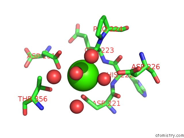
Mono view
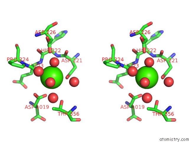
Stereo pair view
Mono view
Stereo pair view
A full contact list of Calcium with other atoms in the Ca binding
site number 1 of Crystal Structure of Human Aminopeptidase A Complexed with Aspartate and Calcium within 5.0Å range:
|
Reference:
Y.Yang,
C.Liu,
Y.L.Lin,
F.Li.
Structural Insights Into Central Hypertension Regulation By Human Aminopeptidase A. J.Biol.Chem. V. 288 25638 2013.
ISSN: ISSN 0021-9258
PubMed: 23888046
DOI: 10.1074/JBC.M113.494955
Page generated: Sun Jul 14 09:20:31 2024
ISSN: ISSN 0021-9258
PubMed: 23888046
DOI: 10.1074/JBC.M113.494955
Last articles
Zn in 9MJ5Zn in 9HNW
Zn in 9G0L
Zn in 9FNE
Zn in 9DZN
Zn in 9E0I
Zn in 9D32
Zn in 9DAK
Zn in 8ZXC
Zn in 8ZUF