Calcium »
PDB 4llt-4m17 »
4lt7 »
Calcium in PDB 4lt7: Crystal Structure of the C2A Domain of Rabphilin-3A in Complex with A Calcium
Protein crystallography data
The structure of Crystal Structure of the C2A Domain of Rabphilin-3A in Complex with A Calcium, PDB code: 4lt7
was solved by
N.Verdaguer,
C Ferrer-Orta,
M.Buxaderas,
S.Corbalan-Garcia,
D.Perez-Sanchez,
M.Guerrero-Valero,
G.Luengo,
J.Pous,
P.Guerra,
J.C.Gomez-Fernandez,
J.Guillen,
with X-Ray Crystallography technique. A brief refinement statistics is given in the table below:
| Resolution Low / High (Å) | 40.00 / 2.50 |
| Space group | P 21 21 21 |
| Cell size a, b, c (Å), α, β, γ (°) | 38.690, 40.030, 90.250, 90.00, 90.00, 90.00 |
| R / Rfree (%) | 24.3 / 28.9 |
Calcium Binding Sites:
The binding sites of Calcium atom in the Crystal Structure of the C2A Domain of Rabphilin-3A in Complex with A Calcium
(pdb code 4lt7). This binding sites where shown within
5.0 Angstroms radius around Calcium atom.
In total only one binding site of Calcium was determined in the Crystal Structure of the C2A Domain of Rabphilin-3A in Complex with A Calcium, PDB code: 4lt7:
In total only one binding site of Calcium was determined in the Crystal Structure of the C2A Domain of Rabphilin-3A in Complex with A Calcium, PDB code: 4lt7:
Calcium binding site 1 out of 1 in 4lt7
Go back to
Calcium binding site 1 out
of 1 in the Crystal Structure of the C2A Domain of Rabphilin-3A in Complex with A Calcium
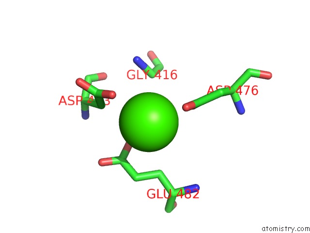
Mono view
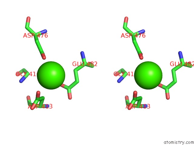
Stereo pair view
Mono view
Stereo pair view
A full contact list of Calcium with other atoms in the Ca binding
site number 1 of Crystal Structure of the C2A Domain of Rabphilin-3A in Complex with A Calcium within 5.0Å range:
|
Reference:
J.Guillen,
C.Ferrer-Orta,
M.Buxaderas,
D.Perez-Sanchez,
M.Guerrero-Valero,
G.Luengo-Gil,
J.Pous,
P.Guerra,
J.C.Gomez-Fernandez,
N.Verdaguer,
S.Corbalan-Garcia.
Structural Insights Into the CA2+ and Pi(4,5)P2 Binding Modes of the C2 Domains of Rabphilin 3A and Synaptotagmin 1. Proc.Natl.Acad.Sci.Usa V. 110 20503 2013.
ISSN: ISSN 0027-8424
PubMed: 24302762
DOI: 10.1073/PNAS.1316179110
Page generated: Tue Jul 8 23:58:36 2025
ISSN: ISSN 0027-8424
PubMed: 24302762
DOI: 10.1073/PNAS.1316179110
Last articles
Cl in 5R90Cl in 5R8Z
Cl in 5R8Y
Cl in 5R8X
Cl in 5R8W
Cl in 5R83
Cl in 5R8V
Cl in 5R8U
Cl in 5R7X
Cl in 5R7Z