Calcium »
PDB 4llt-4m17 »
4lud »
Calcium in PDB 4lud: Crystal Structure of Hck in Complex with the Fluorescent Compound SKF86002
Enzymatic activity of Crystal Structure of Hck in Complex with the Fluorescent Compound SKF86002
All present enzymatic activity of Crystal Structure of Hck in Complex with the Fluorescent Compound SKF86002:
2.7.10.2;
2.7.10.2;
Protein crystallography data
The structure of Crystal Structure of Hck in Complex with the Fluorescent Compound SKF86002, PDB code: 4lud
was solved by
L.J.Parker,
A.Tanaka,
N.Handa,
K.Honda,
Y.Tomabechi,
M.Shirouzu,
S.Yokoyama,
with X-Ray Crystallography technique. A brief refinement statistics is given in the table below:
| Resolution Low / High (Å) | 47.76 / 2.85 |
| Space group | P 1 21 1 |
| Cell size a, b, c (Å), α, β, γ (°) | 48.300, 73.400, 180.290, 90.00, 96.77, 90.00 |
| R / Rfree (%) | 21.2 / 26.1 |
Other elements in 4lud:
The structure of Crystal Structure of Hck in Complex with the Fluorescent Compound SKF86002 also contains other interesting chemical elements:
| Fluorine | (F) | 2 atoms |
| Chlorine | (Cl) | 2 atoms |
Calcium Binding Sites:
The binding sites of Calcium atom in the Crystal Structure of Hck in Complex with the Fluorescent Compound SKF86002
(pdb code 4lud). This binding sites where shown within
5.0 Angstroms radius around Calcium atom.
In total 3 binding sites of Calcium where determined in the Crystal Structure of Hck in Complex with the Fluorescent Compound SKF86002, PDB code: 4lud:
Jump to Calcium binding site number: 1; 2; 3;
In total 3 binding sites of Calcium where determined in the Crystal Structure of Hck in Complex with the Fluorescent Compound SKF86002, PDB code: 4lud:
Jump to Calcium binding site number: 1; 2; 3;
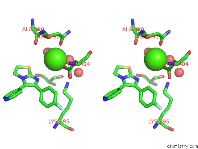
Calcium binding site 1 out of 3 in 4lud
Go back to
Calcium binding site 1 out
of 3 in the Crystal Structure of Hck in Complex with the Fluorescent Compound SKF86002
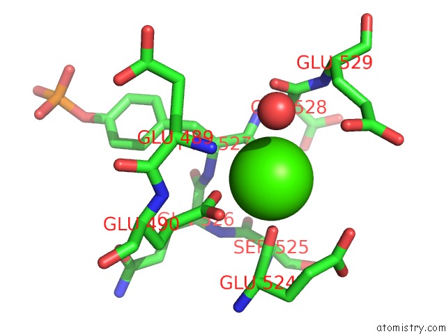
Mono view
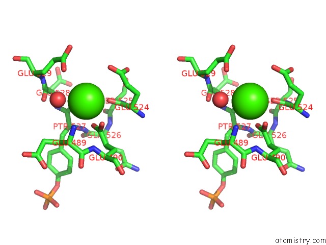
Stereo pair view
Mono view
Stereo pair view
A full contact list of Calcium with other atoms in the Ca binding
site number 1 of Crystal Structure of Hck in Complex with the Fluorescent Compound SKF86002 within 5.0Å range:
|
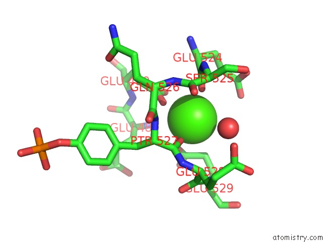
Calcium binding site 2 out of 3 in 4lud
Go back to
Calcium binding site 2 out
of 3 in the Crystal Structure of Hck in Complex with the Fluorescent Compound SKF86002
Mono view

Stereo pair view
Mono view
Stereo pair view
A full contact list of Calcium with other atoms in the Ca binding
site number 2 of Crystal Structure of Hck in Complex with the Fluorescent Compound SKF86002 within 5.0Å range:
|
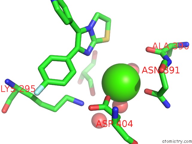
Calcium binding site 3 out of 3 in 4lud
Go back to
Calcium binding site 3 out
of 3 in the Crystal Structure of Hck in Complex with the Fluorescent Compound SKF86002
Mono view
Stereo pair view
Mono view
Stereo pair view
A full contact list of Calcium with other atoms in the Ca binding
site number 3 of Crystal Structure of Hck in Complex with the Fluorescent Compound SKF86002 within 5.0Å range:
|
Reference:
L.J.Parker,
S.Taruya,
K.Tsuganezawa,
N.Ogawa,
J.Mikuni,
K.Honda,
T.Tomabechi,
N.Handa,
M.Shirouzu,
S.Yokoyama,
A.Tanaka.
Kinase Crystal Identification and Atp-Competitive Inhibitor Screening Using the Fluorescent Ligand SKF86002 Acta Crystallogr.,Sect.D V. 70 392 2014.
ISSN: ISSN 0907-4449
DOI: 10.1107/S1399004713028654
Page generated: Sun Jul 14 09:45:25 2024
ISSN: ISSN 0907-4449
DOI: 10.1107/S1399004713028654
Last articles
Zn in 9MJ5Zn in 9HNW
Zn in 9G0L
Zn in 9FNE
Zn in 9DZN
Zn in 9E0I
Zn in 9D32
Zn in 9DAK
Zn in 8ZXC
Zn in 8ZUF