Calcium »
PDB 4m18-4m8u »
4m2q »
Calcium in PDB 4m2q: Crystal Structure of Non-Myristoylated Recoverin with Cysteine-39 Oxidized to Sulfenic Acid
Protein crystallography data
The structure of Crystal Structure of Non-Myristoylated Recoverin with Cysteine-39 Oxidized to Sulfenic Acid, PDB code: 4m2q
was solved by
R.Prem Kumar,
K.Chakrabarti,
D.Kern,
D.D.Oprian,
with X-Ray Crystallography technique. A brief refinement statistics is given in the table below:
| Resolution Low / High (Å) | 42.00 / 1.90 |
| Space group | I 4 |
| Cell size a, b, c (Å), α, β, γ (°) | 84.640, 84.640, 59.480, 90.00, 90.00, 90.00 |
| R / Rfree (%) | 18.9 / 22.1 |
Calcium Binding Sites:
The binding sites of Calcium atom in the Crystal Structure of Non-Myristoylated Recoverin with Cysteine-39 Oxidized to Sulfenic Acid
(pdb code 4m2q). This binding sites where shown within
5.0 Angstroms radius around Calcium atom.
In total only one binding site of Calcium was determined in the Crystal Structure of Non-Myristoylated Recoverin with Cysteine-39 Oxidized to Sulfenic Acid, PDB code: 4m2q:
In total only one binding site of Calcium was determined in the Crystal Structure of Non-Myristoylated Recoverin with Cysteine-39 Oxidized to Sulfenic Acid, PDB code: 4m2q:
Calcium binding site 1 out of 1 in 4m2q
Go back to
Calcium binding site 1 out
of 1 in the Crystal Structure of Non-Myristoylated Recoverin with Cysteine-39 Oxidized to Sulfenic Acid
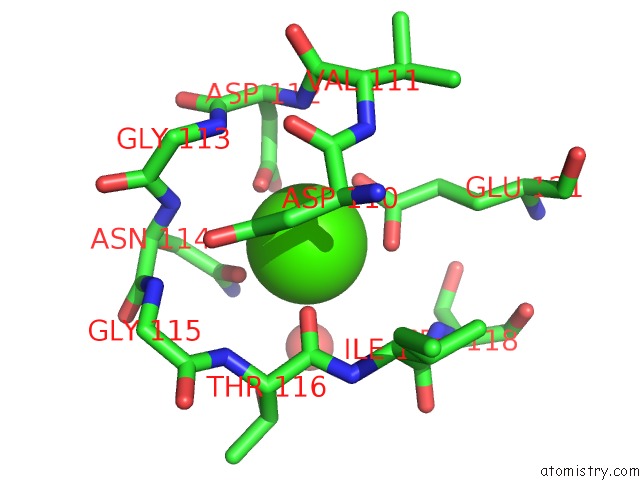
Mono view
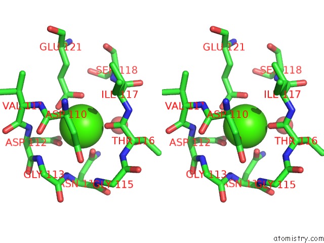
Stereo pair view
Mono view
Stereo pair view
A full contact list of Calcium with other atoms in the Ca binding
site number 1 of Crystal Structure of Non-Myristoylated Recoverin with Cysteine-39 Oxidized to Sulfenic Acid within 5.0Å range:
|
Reference:
M.J.Ranaghan,
R.P.Kumar,
K.S.Chakrabarti,
V.Buosi,
D.Kern,
D.D.Oprian.
A Highly Conserved Cysteine of Neuronal Calcium-Sensing Proteins Controls Cooperative Binding of CA2+ to Recoverin. J.Biol.Chem. V. 288 36160 2013.
ISSN: ISSN 0021-9258
PubMed: 24189072
DOI: 10.1074/JBC.M113.524355
Page generated: Sun Jul 14 09:58:54 2024
ISSN: ISSN 0021-9258
PubMed: 24189072
DOI: 10.1074/JBC.M113.524355
Last articles
Zn in 9J0NZn in 9J0O
Zn in 9J0P
Zn in 9FJX
Zn in 9EKB
Zn in 9C0F
Zn in 9CAH
Zn in 9CH0
Zn in 9CH3
Zn in 9CH1