Calcium »
PDB 4m18-4m8u »
4m7k »
Calcium in PDB 4m7k: Crystal Structure of Anti-Tissue Factor Antibody 10H10
Protein crystallography data
The structure of Crystal Structure of Anti-Tissue Factor Antibody 10H10, PDB code: 4m7k
was solved by
A.Teplyakov,
G.Obmolova,
T.Malia,
G.L.Gilliland,
with X-Ray Crystallography technique. A brief refinement statistics is given in the table below:
| Resolution Low / High (Å) | 15.00 / 1.90 |
| Space group | C 2 2 21 |
| Cell size a, b, c (Å), α, β, γ (°) | 81.770, 135.510, 88.250, 90.00, 90.00, 90.00 |
| R / Rfree (%) | 20.5 / 24.3 |
Calcium Binding Sites:
The binding sites of Calcium atom in the Crystal Structure of Anti-Tissue Factor Antibody 10H10
(pdb code 4m7k). This binding sites where shown within
5.0 Angstroms radius around Calcium atom.
In total only one binding site of Calcium was determined in the Crystal Structure of Anti-Tissue Factor Antibody 10H10, PDB code: 4m7k:
In total only one binding site of Calcium was determined in the Crystal Structure of Anti-Tissue Factor Antibody 10H10, PDB code: 4m7k:
Calcium binding site 1 out of 1 in 4m7k
Go back to
Calcium binding site 1 out
of 1 in the Crystal Structure of Anti-Tissue Factor Antibody 10H10
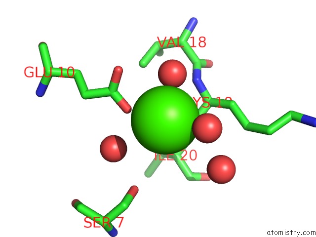
Mono view
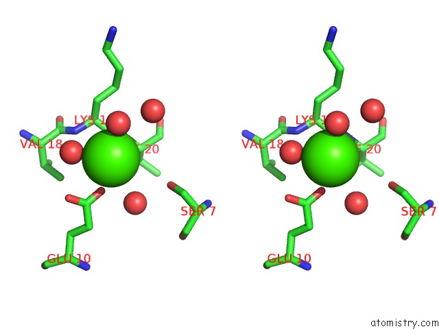
Stereo pair view
Mono view
Stereo pair view
A full contact list of Calcium with other atoms in the Ca binding
site number 1 of Crystal Structure of Anti-Tissue Factor Antibody 10H10 within 5.0Å range:
|
Reference:
A.Teplyakov,
J.Luo,
G.Obmolova,
T.J.Malia,
R.Sweet,
R.L.Stanfield,
S.Kodangattil,
J.C.Almagro,
G.L.Gilliland.
Antibody Modeling Assessment II. Structures and Models. Proteins V. 82 1563 2014.
ISSN: ISSN 0887-3585
PubMed: 24633955
DOI: 10.1002/PROT.24554
Page generated: Sun Jul 14 10:14:45 2024
ISSN: ISSN 0887-3585
PubMed: 24633955
DOI: 10.1002/PROT.24554
Last articles
Zn in 9J0NZn in 9J0O
Zn in 9J0P
Zn in 9FJX
Zn in 9EKB
Zn in 9C0F
Zn in 9CAH
Zn in 9CH0
Zn in 9CH3
Zn in 9CH1