Calcium »
PDB 4m93-4mpp »
4mh1 »
Calcium in PDB 4mh1: Crystal Structure and Functional Studies of Quinoprotein L-Sorbose Dehydrogenase From Ketogulonicigenium Vulgare Y25
Protein crystallography data
The structure of Crystal Structure and Functional Studies of Quinoprotein L-Sorbose Dehydrogenase From Ketogulonicigenium Vulgare Y25, PDB code: 4mh1
was solved by
X.Han,
X.Liu,
with X-Ray Crystallography technique. A brief refinement statistics is given in the table below:
| Resolution Low / High (Å) | 41.14 / 2.70 |
| Space group | P 4 3 2 |
| Cell size a, b, c (Å), α, β, γ (°) | 201.530, 201.530, 201.530, 90.00, 90.00, 90.00 |
| R / Rfree (%) | 22.6 / 23.2 |
Calcium Binding Sites:
The binding sites of Calcium atom in the Crystal Structure and Functional Studies of Quinoprotein L-Sorbose Dehydrogenase From Ketogulonicigenium Vulgare Y25
(pdb code 4mh1). This binding sites where shown within
5.0 Angstroms radius around Calcium atom.
In total only one binding site of Calcium was determined in the Crystal Structure and Functional Studies of Quinoprotein L-Sorbose Dehydrogenase From Ketogulonicigenium Vulgare Y25, PDB code: 4mh1:
In total only one binding site of Calcium was determined in the Crystal Structure and Functional Studies of Quinoprotein L-Sorbose Dehydrogenase From Ketogulonicigenium Vulgare Y25, PDB code: 4mh1:
Calcium binding site 1 out of 1 in 4mh1
Go back to
Calcium binding site 1 out
of 1 in the Crystal Structure and Functional Studies of Quinoprotein L-Sorbose Dehydrogenase From Ketogulonicigenium Vulgare Y25
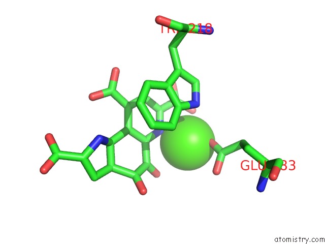
Mono view
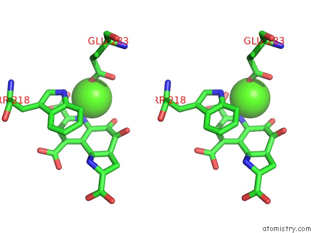
Stereo pair view
Mono view
Stereo pair view
A full contact list of Calcium with other atoms in the Ca binding
site number 1 of Crystal Structure and Functional Studies of Quinoprotein L-Sorbose Dehydrogenase From Ketogulonicigenium Vulgare Y25 within 5.0Å range:
|
Reference:
X.Han,
X.Xiong,
D.Jiang,
S.Chen,
E.Huang,
W.Zhang,
X.Liu.
Crystal Structure of L-Sorbose Dehydrogenase, A Pyrroloquinoline Quinone-Dependent Enzyme with Homodimeric Assembly, From Ketogulonicigenium Vulgare Biotechnol.Lett. V. 36 1001 2014.
ISSN: ISSN 0141-5492
PubMed: 24557074
DOI: 10.1007/S10529-013-1446-5
Page generated: Sun Jul 14 10:22:46 2024
ISSN: ISSN 0141-5492
PubMed: 24557074
DOI: 10.1007/S10529-013-1446-5
Last articles
Zn in 9J0NZn in 9J0O
Zn in 9J0P
Zn in 9FJX
Zn in 9EKB
Zn in 9C0F
Zn in 9CAH
Zn in 9CH0
Zn in 9CH3
Zn in 9CH1