Calcium »
PDB 4m93-4mpp »
4mmh »
Calcium in PDB 4mmh: Crystal Structure of Heparan Sulfate Lyase Hepc From Pedobacter Heparinus
Enzymatic activity of Crystal Structure of Heparan Sulfate Lyase Hepc From Pedobacter Heparinus
All present enzymatic activity of Crystal Structure of Heparan Sulfate Lyase Hepc From Pedobacter Heparinus:
4.2.2.8;
4.2.2.8;
Protein crystallography data
The structure of Crystal Structure of Heparan Sulfate Lyase Hepc From Pedobacter Heparinus, PDB code: 4mmh
was solved by
Y.Maruyama,
Y.Nakamichi,
B.Mikami,
K.Murata,
W.Hashimoto,
with X-Ray Crystallography technique. A brief refinement statistics is given in the table below:
| Resolution Low / High (Å) | 39.67 / 2.20 |
| Space group | P 21 21 21 |
| Cell size a, b, c (Å), α, β, γ (°) | 41.245, 104.067, 143.307, 90.00, 90.00, 90.00 |
| R / Rfree (%) | 19.5 / 24.4 |
Calcium Binding Sites:
The binding sites of Calcium atom in the Crystal Structure of Heparan Sulfate Lyase Hepc From Pedobacter Heparinus
(pdb code 4mmh). This binding sites where shown within
5.0 Angstroms radius around Calcium atom.
In total 2 binding sites of Calcium where determined in the Crystal Structure of Heparan Sulfate Lyase Hepc From Pedobacter Heparinus, PDB code: 4mmh:
Jump to Calcium binding site number: 1; 2;
In total 2 binding sites of Calcium where determined in the Crystal Structure of Heparan Sulfate Lyase Hepc From Pedobacter Heparinus, PDB code: 4mmh:
Jump to Calcium binding site number: 1; 2;
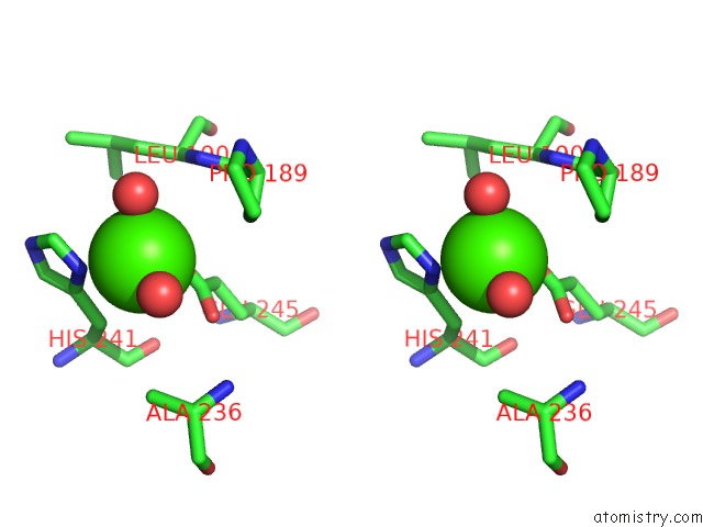
Calcium binding site 1 out of 2 in 4mmh
Go back to
Calcium binding site 1 out
of 2 in the Crystal Structure of Heparan Sulfate Lyase Hepc From Pedobacter Heparinus

Mono view
Stereo pair view
Mono view
Stereo pair view
A full contact list of Calcium with other atoms in the Ca binding
site number 1 of Crystal Structure of Heparan Sulfate Lyase Hepc From Pedobacter Heparinus within 5.0Å range:
|
Calcium binding site 2 out of 2 in 4mmh
Go back to
Calcium binding site 2 out
of 2 in the Crystal Structure of Heparan Sulfate Lyase Hepc From Pedobacter Heparinus

Mono view

Stereo pair view
Mono view
Stereo pair view
A full contact list of Calcium with other atoms in the Ca binding
site number 2 of Crystal Structure of Heparan Sulfate Lyase Hepc From Pedobacter Heparinus within 5.0Å range:
|
Reference:
W.Hashimoto,
Y.Maruyama,
Y.Nakamichi,
B.Mikami,
K.Murata.
Crystal Structure of Pedobacter Heparinus Heparin Lyase Hep III with the Active Site in A Deep Cleft Biochemistry V. 53 777 2014.
ISSN: ISSN 0006-2960
PubMed: 24437462
DOI: 10.1021/BI4012463
Page generated: Sun Jul 14 10:27:37 2024
ISSN: ISSN 0006-2960
PubMed: 24437462
DOI: 10.1021/BI4012463
Last articles
Zn in 9J0NZn in 9J0O
Zn in 9J0P
Zn in 9FJX
Zn in 9EKB
Zn in 9C0F
Zn in 9CAH
Zn in 9CH0
Zn in 9CH3
Zn in 9CH1