Calcium »
PDB 4m93-4mpp »
4mn0 »
Calcium in PDB 4mn0: Spatial Structure of the Novel Light-Sensitive Photoprotein Berovin From the Ctenophore Beroe Abyssicola in the CA2+-Loaded Apoprotein Conformation State
Protein crystallography data
The structure of Spatial Structure of the Novel Light-Sensitive Photoprotein Berovin From the Ctenophore Beroe Abyssicola in the CA2+-Loaded Apoprotein Conformation State, PDB code: 4mn0
was solved by
Z.J.Liu,
G.A.Stepanyuk,
E.S.Vysotski,
J.Lee,
J.P.Rose,
B.C.Wang,
Southeastcollaboratory For Structural Genomics (Secsg),
with X-Ray Crystallography technique. A brief refinement statistics is given in the table below:
| Resolution Low / High (Å) | 40.78 / 1.90 |
| Space group | C 1 2 1 |
| Cell size a, b, c (Å), α, β, γ (°) | 101.770, 33.899, 77.406, 90.00, 126.74, 90.00 |
| R / Rfree (%) | 19.3 / 25.6 |
Other elements in 4mn0:
The structure of Spatial Structure of the Novel Light-Sensitive Photoprotein Berovin From the Ctenophore Beroe Abyssicola in the CA2+-Loaded Apoprotein Conformation State also contains other interesting chemical elements:
| Magnesium | (Mg) | 1 atom |
Calcium Binding Sites:
The binding sites of Calcium atom in the Spatial Structure of the Novel Light-Sensitive Photoprotein Berovin From the Ctenophore Beroe Abyssicola in the CA2+-Loaded Apoprotein Conformation State
(pdb code 4mn0). This binding sites where shown within
5.0 Angstroms radius around Calcium atom.
In total 3 binding sites of Calcium where determined in the Spatial Structure of the Novel Light-Sensitive Photoprotein Berovin From the Ctenophore Beroe Abyssicola in the CA2+-Loaded Apoprotein Conformation State, PDB code: 4mn0:
Jump to Calcium binding site number: 1; 2; 3;
In total 3 binding sites of Calcium where determined in the Spatial Structure of the Novel Light-Sensitive Photoprotein Berovin From the Ctenophore Beroe Abyssicola in the CA2+-Loaded Apoprotein Conformation State, PDB code: 4mn0:
Jump to Calcium binding site number: 1; 2; 3;
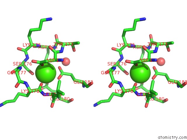
Calcium binding site 1 out of 3 in 4mn0
Go back to
Calcium binding site 1 out
of 3 in the Spatial Structure of the Novel Light-Sensitive Photoprotein Berovin From the Ctenophore Beroe Abyssicola in the CA2+-Loaded Apoprotein Conformation State

Mono view
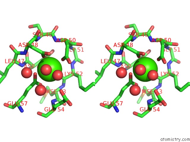
Stereo pair view
Mono view
Stereo pair view
A full contact list of Calcium with other atoms in the Ca binding
site number 1 of Spatial Structure of the Novel Light-Sensitive Photoprotein Berovin From the Ctenophore Beroe Abyssicola in the CA2+-Loaded Apoprotein Conformation State within 5.0Å range:
|
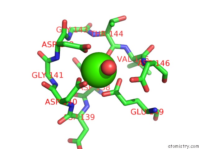
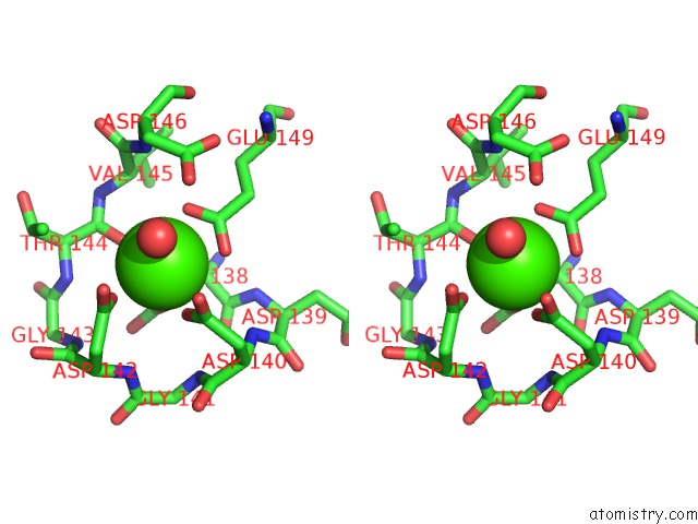
Calcium binding site 2 out of 3 in 4mn0
Go back to
Calcium binding site 2 out
of 3 in the Spatial Structure of the Novel Light-Sensitive Photoprotein Berovin From the Ctenophore Beroe Abyssicola in the CA2+-Loaded Apoprotein Conformation State
Mono view
Stereo pair view
Mono view
Stereo pair view
A full contact list of Calcium with other atoms in the Ca binding
site number 2 of Spatial Structure of the Novel Light-Sensitive Photoprotein Berovin From the Ctenophore Beroe Abyssicola in the CA2+-Loaded Apoprotein Conformation State within 5.0Å range:
|
Calcium binding site 3 out of 3 in 4mn0
Go back to
Calcium binding site 3 out
of 3 in the Spatial Structure of the Novel Light-Sensitive Photoprotein Berovin From the Ctenophore Beroe Abyssicola in the CA2+-Loaded Apoprotein Conformation State
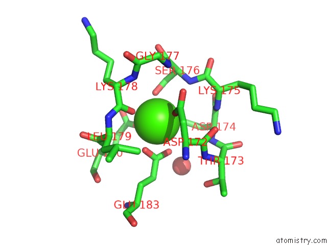
Mono view
Stereo pair view
Mono view
Stereo pair view
A full contact list of Calcium with other atoms in the Ca binding
site number 3 of Spatial Structure of the Novel Light-Sensitive Photoprotein Berovin From the Ctenophore Beroe Abyssicola in the CA2+-Loaded Apoprotein Conformation State within 5.0Å range:
|
Reference:
G.A.Stepanyuk,
Z.J.Liu,
L.P.Burakova,
J.Lee,
J.Rose,
E.S.Vysotski,
B.C.Wang.
Spatial Structure of the Novel Light-Sensitive Photoprotein Berovin From the Ctenophore Beroe Abyssicola in the Ca(2+)-Loaded Apoprotein Conformation State. Biochim.Biophys.Acta V.1834 2139 2013.
ISSN: ISSN 0006-3002
PubMed: 23891746
DOI: 10.1016/J.BBAPAP.2013.07.006
Page generated: Sun Jul 14 10:28:39 2024
ISSN: ISSN 0006-3002
PubMed: 23891746
DOI: 10.1016/J.BBAPAP.2013.07.006
Last articles
Zn in 9MJ5Zn in 9HNW
Zn in 9G0L
Zn in 9FNE
Zn in 9DZN
Zn in 9E0I
Zn in 9D32
Zn in 9DAK
Zn in 8ZXC
Zn in 8ZUF