Calcium »
PDB 4n0b-4n5x »
4n2z »
Calcium in PDB 4n2z: Crystal Structure of the Alpha-L-Arabinofuranosidase PAABF62A From Podospora Anserina in Complex with Cellotriose
Enzymatic activity of Crystal Structure of the Alpha-L-Arabinofuranosidase PAABF62A From Podospora Anserina in Complex with Cellotriose
All present enzymatic activity of Crystal Structure of the Alpha-L-Arabinofuranosidase PAABF62A From Podospora Anserina in Complex with Cellotriose:
3.2.1.55;
3.2.1.55;
Protein crystallography data
The structure of Crystal Structure of the Alpha-L-Arabinofuranosidase PAABF62A From Podospora Anserina in Complex with Cellotriose, PDB code: 4n2z
was solved by
B.Siguier,
C.Dumon,
L.Mourey,
S.Tranier,
with X-Ray Crystallography technique. A brief refinement statistics is given in the table below:
| Resolution Low / High (Å) | 14.84 / 1.80 |
| Space group | C 1 2 1 |
| Cell size a, b, c (Å), α, β, γ (°) | 102.140, 66.790, 60.360, 90.00, 117.33, 90.00 |
| R / Rfree (%) | 13.1 / 17 |
Calcium Binding Sites:
The binding sites of Calcium atom in the Crystal Structure of the Alpha-L-Arabinofuranosidase PAABF62A From Podospora Anserina in Complex with Cellotriose
(pdb code 4n2z). This binding sites where shown within
5.0 Angstroms radius around Calcium atom.
In total only one binding site of Calcium was determined in the Crystal Structure of the Alpha-L-Arabinofuranosidase PAABF62A From Podospora Anserina in Complex with Cellotriose, PDB code: 4n2z:
In total only one binding site of Calcium was determined in the Crystal Structure of the Alpha-L-Arabinofuranosidase PAABF62A From Podospora Anserina in Complex with Cellotriose, PDB code: 4n2z:
Calcium binding site 1 out of 1 in 4n2z
Go back to
Calcium binding site 1 out
of 1 in the Crystal Structure of the Alpha-L-Arabinofuranosidase PAABF62A From Podospora Anserina in Complex with Cellotriose
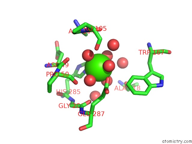
Mono view
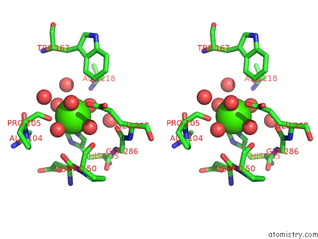
Stereo pair view
Mono view
Stereo pair view
A full contact list of Calcium with other atoms in the Ca binding
site number 1 of Crystal Structure of the Alpha-L-Arabinofuranosidase PAABF62A From Podospora Anserina in Complex with Cellotriose within 5.0Å range:
|
Reference:
B.Siguier,
M.Haon,
V.Nahoum,
M.Marcellin,
O.Burlet-Schiltz,
P.M.Coutinho,
B.Henrissat,
L.Mourey,
M.J.O'donohue,
J.G.Berrin,
S.Tranier,
C.Dumon.
First Structural Insights Into Alpha-L-Arabinofuranosidases From the Two GH62 Glycoside Hydrolase Subfamilies. J.Biol.Chem. V. 289 5261 2014.
ISSN: ISSN 0021-9258
PubMed: 24394409
DOI: 10.1074/JBC.M113.528133
Page generated: Sun Jul 14 10:49:23 2024
ISSN: ISSN 0021-9258
PubMed: 24394409
DOI: 10.1074/JBC.M113.528133
Last articles
Zn in 9J0NZn in 9J0O
Zn in 9J0P
Zn in 9FJX
Zn in 9EKB
Zn in 9C0F
Zn in 9CAH
Zn in 9CH0
Zn in 9CH3
Zn in 9CH1