Calcium »
PDB 4n66-4nhf »
4ncy »
Calcium in PDB 4ncy: In Situ Trypsin Crystallized on A Mitegen Micromesh with Imidazole Ligand
Enzymatic activity of In Situ Trypsin Crystallized on A Mitegen Micromesh with Imidazole Ligand
All present enzymatic activity of In Situ Trypsin Crystallized on A Mitegen Micromesh with Imidazole Ligand:
3.4.21.4;
3.4.21.4;
Protein crystallography data
The structure of In Situ Trypsin Crystallized on A Mitegen Micromesh with Imidazole Ligand, PDB code: 4ncy
was solved by
X.Yin,
A.Scalia,
L.Leroy,
C.M.Cuttitta,
G.M.Polizzo,
D.L.Ericson,
C.G.Roessler,
O.Campos,
R.Agarwal,
M.Allaire,
A.M.Orville,
R.Jackimowicz,
M.Y.Ma,
R.M.Sweet,
A.S.Soares,
with X-Ray Crystallography technique. A brief refinement statistics is given in the table below:
| Resolution Low / High (Å) | 43.77 / 1.42 |
| Space group | P 21 21 21 |
| Cell size a, b, c (Å), α, β, γ (°) | 54.392, 58.186, 66.430, 90.00, 90.00, 90.00 |
| R / Rfree (%) | 11.8 / 13.8 |
Calcium Binding Sites:
The binding sites of Calcium atom in the In Situ Trypsin Crystallized on A Mitegen Micromesh with Imidazole Ligand
(pdb code 4ncy). This binding sites where shown within
5.0 Angstroms radius around Calcium atom.
In total only one binding site of Calcium was determined in the In Situ Trypsin Crystallized on A Mitegen Micromesh with Imidazole Ligand, PDB code: 4ncy:
In total only one binding site of Calcium was determined in the In Situ Trypsin Crystallized on A Mitegen Micromesh with Imidazole Ligand, PDB code: 4ncy:
Calcium binding site 1 out of 1 in 4ncy
Go back to
Calcium binding site 1 out
of 1 in the In Situ Trypsin Crystallized on A Mitegen Micromesh with Imidazole Ligand
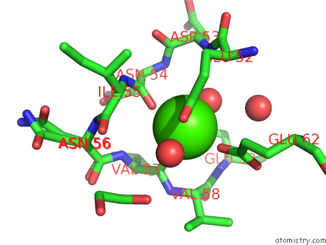
Mono view
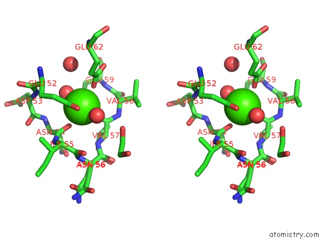
Stereo pair view
Mono view
Stereo pair view
A full contact list of Calcium with other atoms in the Ca binding
site number 1 of In Situ Trypsin Crystallized on A Mitegen Micromesh with Imidazole Ligand within 5.0Å range:
|
Reference:
X.Yin,
A.Scalia,
L.Leroy,
C.M.Cuttitta,
G.M.Polizzo,
D.L.Ericson,
C.G.Roessler,
O.Campos,
M.Y.Ma,
R.Agarwal,
R.Jackimowicz,
M.Allaire,
A.M.Orville,
R.M.Sweet,
A.S.Soares.
Hitting the Target: Fragment Screening with Acoustic in Situ Co-Crystallization of Proteins Plus Fragment Libraries on Pin-Mounted Data-Collection Micromeshes. Acta Crystallogr.,Sect.D V. 70 1177 2014.
ISSN: ISSN 0907-4449
PubMed: 24816088
DOI: 10.1107/S1399004713034603
Page generated: Wed Jul 9 00:48:20 2025
ISSN: ISSN 0907-4449
PubMed: 24816088
DOI: 10.1107/S1399004713034603
Last articles
Fe in 2YXOFe in 2YRS
Fe in 2YXC
Fe in 2YNM
Fe in 2YVJ
Fe in 2YP1
Fe in 2YU2
Fe in 2YU1
Fe in 2YQB
Fe in 2YOO