Calcium »
PDB 4niv-4o4h »
4o03 »
Calcium in PDB 4o03: Crystal Structure of CA2+ Bound Prothrombin Deletion Mutant Residues 146-167
Enzymatic activity of Crystal Structure of CA2+ Bound Prothrombin Deletion Mutant Residues 146-167
All present enzymatic activity of Crystal Structure of CA2+ Bound Prothrombin Deletion Mutant Residues 146-167:
3.4.21.5;
3.4.21.5;
Protein crystallography data
The structure of Crystal Structure of CA2+ Bound Prothrombin Deletion Mutant Residues 146-167, PDB code: 4o03
was solved by
N.Pozzi,
Z.Chen,
D.B.Shropshire,
L.A.Pelc,
E.Di Cera,
with X-Ray Crystallography technique. A brief refinement statistics is given in the table below:
| Resolution Low / High (Å) | 39.75 / 3.38 |
| Space group | C 1 2 1 |
| Cell size a, b, c (Å), α, β, γ (°) | 177.459, 89.431, 87.982, 90.00, 116.36, 90.00 |
| R / Rfree (%) | 23.3 / 27.9 |
Calcium Binding Sites:
The binding sites of Calcium atom in the Crystal Structure of CA2+ Bound Prothrombin Deletion Mutant Residues 146-167
(pdb code 4o03). This binding sites where shown within
5.0 Angstroms radius around Calcium atom.
In total 5 binding sites of Calcium where determined in the Crystal Structure of CA2+ Bound Prothrombin Deletion Mutant Residues 146-167, PDB code: 4o03:
Jump to Calcium binding site number: 1; 2; 3; 4; 5;
In total 5 binding sites of Calcium where determined in the Crystal Structure of CA2+ Bound Prothrombin Deletion Mutant Residues 146-167, PDB code: 4o03:
Jump to Calcium binding site number: 1; 2; 3; 4; 5;
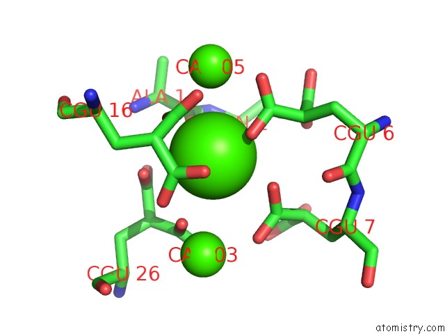
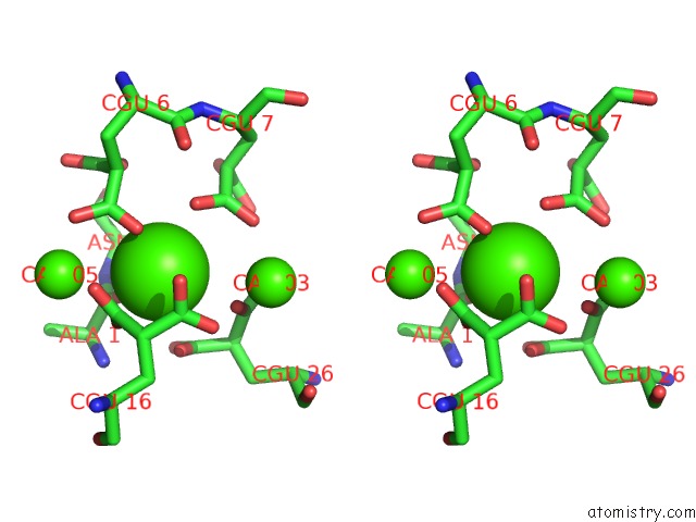
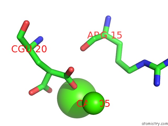
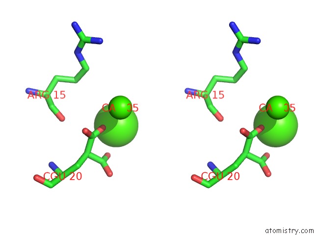
Calcium binding site 1 out of 5 in 4o03
Go back to
Calcium binding site 1 out
of 5 in the Crystal Structure of CA2+ Bound Prothrombin Deletion Mutant Residues 146-167
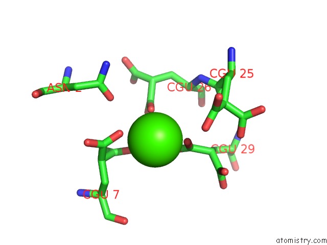
Mono view
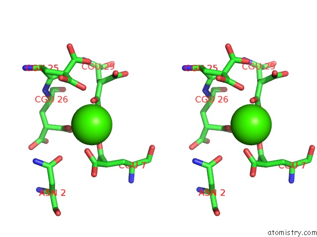
Stereo pair view
Mono view
Stereo pair view
A full contact list of Calcium with other atoms in the Ca binding
site number 1 of Crystal Structure of CA2+ Bound Prothrombin Deletion Mutant Residues 146-167 within 5.0Å range:
|
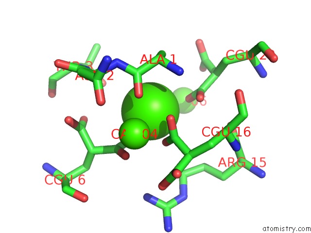
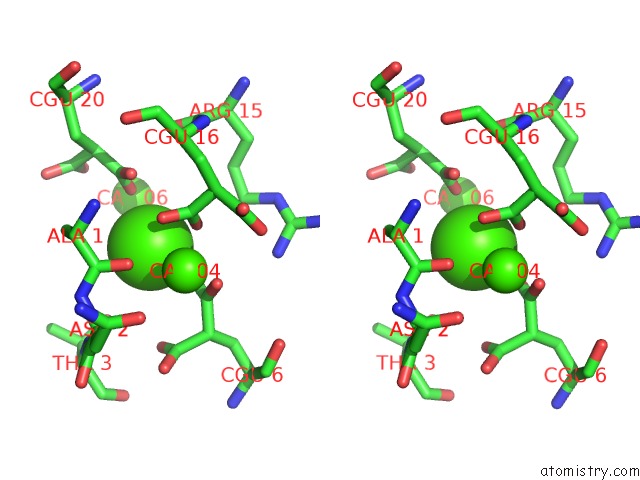
Calcium binding site 2 out of 5 in 4o03
Go back to
Calcium binding site 2 out
of 5 in the Crystal Structure of CA2+ Bound Prothrombin Deletion Mutant Residues 146-167
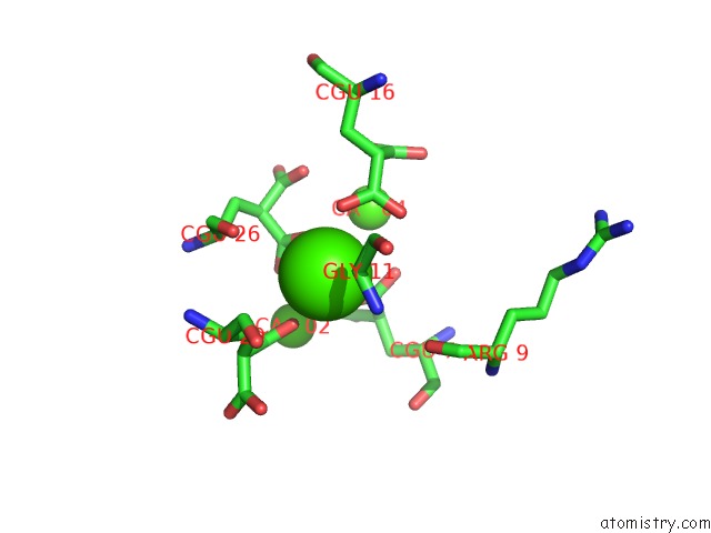
Mono view
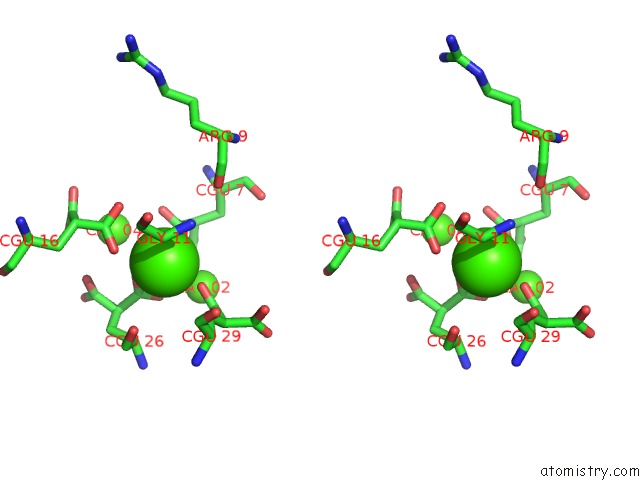
Stereo pair view
Mono view
Stereo pair view
A full contact list of Calcium with other atoms in the Ca binding
site number 2 of Crystal Structure of CA2+ Bound Prothrombin Deletion Mutant Residues 146-167 within 5.0Å range:
|
Calcium binding site 3 out of 5 in 4o03
Go back to
Calcium binding site 3 out
of 5 in the Crystal Structure of CA2+ Bound Prothrombin Deletion Mutant Residues 146-167
Mono view
Stereo pair view
Mono view
Stereo pair view
A full contact list of Calcium with other atoms in the Ca binding
site number 3 of Crystal Structure of CA2+ Bound Prothrombin Deletion Mutant Residues 146-167 within 5.0Å range:
|
Calcium binding site 4 out of 5 in 4o03
Go back to
Calcium binding site 4 out
of 5 in the Crystal Structure of CA2+ Bound Prothrombin Deletion Mutant Residues 146-167
Mono view
Stereo pair view
Mono view
Stereo pair view
A full contact list of Calcium with other atoms in the Ca binding
site number 4 of Crystal Structure of CA2+ Bound Prothrombin Deletion Mutant Residues 146-167 within 5.0Å range:
|
Calcium binding site 5 out of 5 in 4o03
Go back to
Calcium binding site 5 out
of 5 in the Crystal Structure of CA2+ Bound Prothrombin Deletion Mutant Residues 146-167
Mono view
Stereo pair view
Mono view
Stereo pair view
A full contact list of Calcium with other atoms in the Ca binding
site number 5 of Crystal Structure of CA2+ Bound Prothrombin Deletion Mutant Residues 146-167 within 5.0Å range:
|
Reference:
N.Pozzi,
Z.Chen,
L.A.Pelc,
D.B.Shropshire,
E.Di Cera.
The Linker Connecting the Two Kringles Plays A Key Role in Prothrombin Activation. Proc.Natl.Acad.Sci.Usa V. 111 7630 2014.
ISSN: ISSN 0027-8424
PubMed: 24821807
DOI: 10.1073/PNAS.1403779111
Page generated: Sun Jul 14 11:16:40 2024
ISSN: ISSN 0027-8424
PubMed: 24821807
DOI: 10.1073/PNAS.1403779111
Last articles
Zn in 9MJ5Zn in 9HNW
Zn in 9G0L
Zn in 9FNE
Zn in 9DZN
Zn in 9E0I
Zn in 9D32
Zn in 9DAK
Zn in 8ZXC
Zn in 8ZUF