Calcium »
PDB 4o4i-4op8 »
4oj4 »
Calcium in PDB 4oj4: Crystal Structure of V290M Ppargamma Mutant in Complex with Diclofenac
Protein crystallography data
The structure of Crystal Structure of V290M Ppargamma Mutant in Complex with Diclofenac, PDB code: 4oj4
was solved by
A.C.Puhl,
P.Webb,
I.Polikarpov,
with X-Ray Crystallography technique. A brief refinement statistics is given in the table below:
| Resolution Low / High (Å) | 48.19 / 2.30 |
| Space group | P 43 21 2 |
| Cell size a, b, c (Å), α, β, γ (°) | 60.040, 60.040, 161.190, 90.00, 90.00, 90.00 |
| R / Rfree (%) | 21.9 / 28.4 |
Other elements in 4oj4:
The structure of Crystal Structure of V290M Ppargamma Mutant in Complex with Diclofenac also contains other interesting chemical elements:
| Chlorine | (Cl) | 2 atoms |
Calcium Binding Sites:
The binding sites of Calcium atom in the Crystal Structure of V290M Ppargamma Mutant in Complex with Diclofenac
(pdb code 4oj4). This binding sites where shown within
5.0 Angstroms radius around Calcium atom.
In total 2 binding sites of Calcium where determined in the Crystal Structure of V290M Ppargamma Mutant in Complex with Diclofenac, PDB code: 4oj4:
Jump to Calcium binding site number: 1; 2;
In total 2 binding sites of Calcium where determined in the Crystal Structure of V290M Ppargamma Mutant in Complex with Diclofenac, PDB code: 4oj4:
Jump to Calcium binding site number: 1; 2;
Calcium binding site 1 out of 2 in 4oj4
Go back to
Calcium binding site 1 out
of 2 in the Crystal Structure of V290M Ppargamma Mutant in Complex with Diclofenac
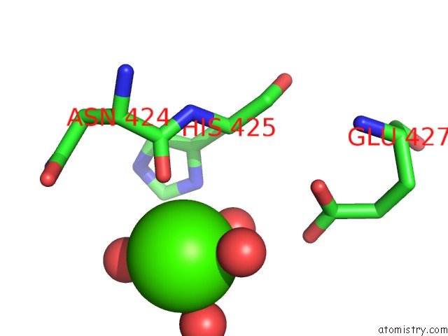
Mono view
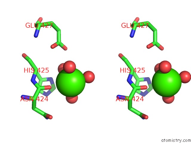
Stereo pair view
Mono view
Stereo pair view
A full contact list of Calcium with other atoms in the Ca binding
site number 1 of Crystal Structure of V290M Ppargamma Mutant in Complex with Diclofenac within 5.0Å range:
|
Calcium binding site 2 out of 2 in 4oj4
Go back to
Calcium binding site 2 out
of 2 in the Crystal Structure of V290M Ppargamma Mutant in Complex with Diclofenac
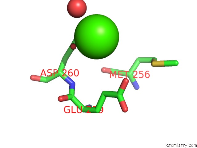
Mono view
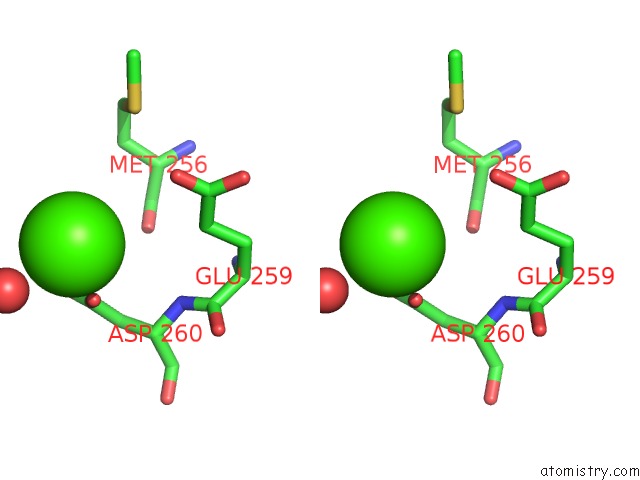
Stereo pair view
Mono view
Stereo pair view
A full contact list of Calcium with other atoms in the Ca binding
site number 2 of Crystal Structure of V290M Ppargamma Mutant in Complex with Diclofenac within 5.0Å range:
|
Reference:
A.C.Puhl,
A.Bernardes,
I.Polikarpov,
P.Webb.
Crystal Structure of A Ppar Ligand-Resistance Syndrome Mutant To Be Published.
Page generated: Wed Jul 9 01:03:08 2025
Last articles
Cl in 5RA4Cl in 5RA6
Cl in 5RA5
Cl in 5RA3
Cl in 5RA2
Cl in 5RA1
Cl in 5R9Z
Cl in 5RA0
Cl in 5R9Y
Cl in 5R9X