Calcium »
PDB 4opq-4p4f »
4opq »
Calcium in PDB 4opq: Room Temperature Crystal Structure of Stabilized Tem-1 Beta-Lactamase Variant V.13 Carrying R164S/G238S Mutations
Protein crystallography data
The structure of Room Temperature Crystal Structure of Stabilized Tem-1 Beta-Lactamase Variant V.13 Carrying R164S/G238S Mutations, PDB code: 4opq
was solved by
E.Dellus-Gur,
M.Elias,
J.S.Fraser,
D.S.Tawfik,
with X-Ray Crystallography technique. A brief refinement statistics is given in the table below:
| Resolution Low / High (Å) | 45.15 / 1.70 |
| Space group | C 1 2 1 |
| Cell size a, b, c (Å), α, β, γ (°) | 154.900, 47.200, 34.890, 90.00, 92.82, 90.00 |
| R / Rfree (%) | 14.9 / 18.9 |
Calcium Binding Sites:
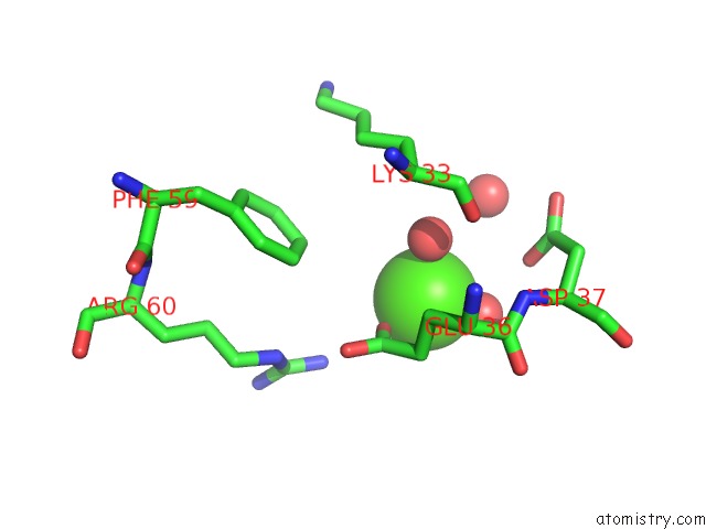
The binding sites of Calcium atom in the Room Temperature Crystal Structure of Stabilized Tem-1 Beta-Lactamase Variant V.13 Carrying R164S/G238S Mutations
(pdb code 4opq). This binding sites where shown within
5.0 Angstroms radius around Calcium atom.
In total only one binding site of Calcium was determined in the Room Temperature Crystal Structure of Stabilized Tem-1 Beta-Lactamase Variant V.13 Carrying R164S/G238S Mutations, PDB code: 4opq:
In total only one binding site of Calcium was determined in the Room Temperature Crystal Structure of Stabilized Tem-1 Beta-Lactamase Variant V.13 Carrying R164S/G238S Mutations, PDB code: 4opq:
Calcium binding site 1 out of 1 in 4opq
Go back to
Calcium binding site 1 out
of 1 in the Room Temperature Crystal Structure of Stabilized Tem-1 Beta-Lactamase Variant V.13 Carrying R164S/G238S Mutations
Mono view
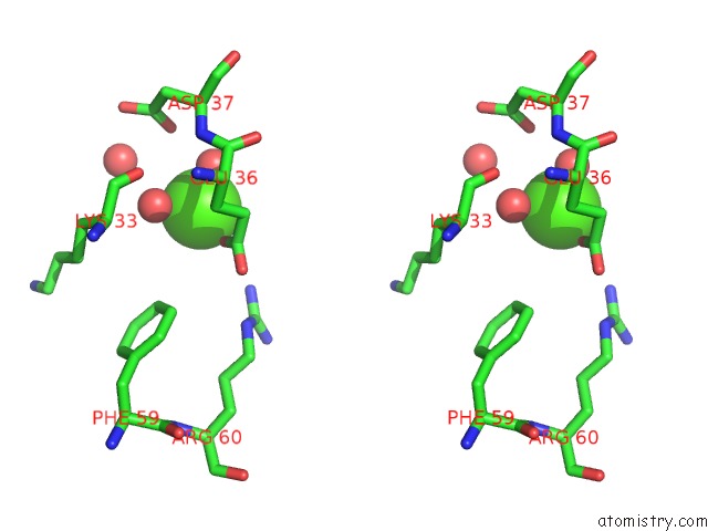
Stereo pair view
Mono view
Stereo pair view
A full contact list of Calcium with other atoms in the Ca binding
site number 1 of Room Temperature Crystal Structure of Stabilized Tem-1 Beta-Lactamase Variant V.13 Carrying R164S/G238S Mutations within 5.0Å range:
|
Reference:
E.Dellus-Gur,
M.Elias,
E.Caselli,
F.Prati,
J.S.Fraser,
D.S.Tawfik.
Negative Epistasis in Enzyme Evolution the Thin Line Between Conformational Freedom and Anarchy To Be Published.
Page generated: Wed Jul 9 01:08:19 2025
Last articles
F in 4IGHF in 4IGA
F in 4IBJ
F in 4IFY
F in 4IFV
F in 4IDO
F in 4ICC
F in 4IBI
F in 4IAH
F in 4IAE