Calcium »
PDB 4opq-4p4f »
4ove »
Calcium in PDB 4ove: X-Ray Crystal Structure of Mouse Netrin-1
Protein crystallography data
The structure of X-Ray Crystal Structure of Mouse Netrin-1, PDB code: 4ove
was solved by
M.Meier,
D.Nikodemus,
R.Reuten,
T.R.Patel,
G.Orriss,
N.Okun,
M.Koch,
J.Stetefeld,
with X-Ray Crystallography technique. A brief refinement statistics is given in the table below:
| Resolution Low / High (Å) | 50.00 / 2.64 |
| Space group | P 32 2 1 |
| Cell size a, b, c (Å), α, β, γ (°) | 69.754, 69.754, 334.802, 90.00, 90.00, 120.00 |
| R / Rfree (%) | 23 / 28.8 |
Other elements in 4ove:
The structure of X-Ray Crystal Structure of Mouse Netrin-1 also contains other interesting chemical elements:
| Chlorine | (Cl) | 1 atom |
Calcium Binding Sites:
The binding sites of Calcium atom in the X-Ray Crystal Structure of Mouse Netrin-1
(pdb code 4ove). This binding sites where shown within
5.0 Angstroms radius around Calcium atom.
In total only one binding site of Calcium was determined in the X-Ray Crystal Structure of Mouse Netrin-1, PDB code: 4ove:
In total only one binding site of Calcium was determined in the X-Ray Crystal Structure of Mouse Netrin-1, PDB code: 4ove:
Calcium binding site 1 out of 1 in 4ove
Go back to
Calcium binding site 1 out
of 1 in the X-Ray Crystal Structure of Mouse Netrin-1

Mono view
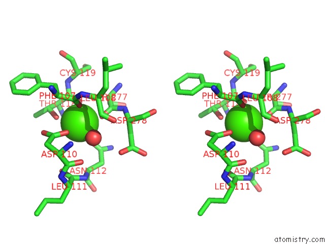
Stereo pair view
Mono view
Stereo pair view
A full contact list of Calcium with other atoms in the Ca binding
site number 1 of X-Ray Crystal Structure of Mouse Netrin-1 within 5.0Å range:
|
Reference:
M.Meier,
D.Nikodemus,
R.Reuten,
T.R.Patel,
G.Orriss,
N.Okun,
K.Mceleney,
F.Schneiders,
K.Poole,
M.Koch,
J.Stetefeld.
Dependence Receptor Recognition By Netrin-1 To Be Published.
Page generated: Wed Jul 9 01:10:02 2025
Last articles
Cl in 5JH2Cl in 5JGZ
Cl in 5JFY
Cl in 5JGN
Cl in 5JGR
Cl in 5JGU
Cl in 5JG1
Cl in 5JGM
Cl in 5JG9
Cl in 5JFF