Calcium »
PDB 4opq-4p4f »
4oy7 »
Calcium in PDB 4oy7: Structure of Cellulose Active Lpmo CELS2 (SCLPMO10C) in Complex with Copper.
Protein crystallography data
The structure of Structure of Cellulose Active Lpmo CELS2 (SCLPMO10C) in Complex with Copper., PDB code: 4oy7
was solved by
Z.Forsberg,
A.K.Mackenzie,
M.Sorlie,
A.K.Rohr,
R.Helland,
A.S.Arvai,
G.Vaaje-Kolstad,
V.G.H.Eijsink,
with X-Ray Crystallography technique. A brief refinement statistics is given in the table below:
| Resolution Low / High (Å) | 47.90 / 1.50 |
| Space group | P 21 21 21 |
| Cell size a, b, c (Å), α, β, γ (°) | 83.858, 122.856, 156.000, 90.00, 90.00, 90.00 |
| R / Rfree (%) | 19.4 / 22.2 |
Other elements in 4oy7:
The structure of Structure of Cellulose Active Lpmo CELS2 (SCLPMO10C) in Complex with Copper. also contains other interesting chemical elements:
| Copper | (Cu) | 8 atoms |
Calcium Binding Sites:
The binding sites of Calcium atom in the Structure of Cellulose Active Lpmo CELS2 (SCLPMO10C) in Complex with Copper.
(pdb code 4oy7). This binding sites where shown within
5.0 Angstroms radius around Calcium atom.
In total 4 binding sites of Calcium where determined in the Structure of Cellulose Active Lpmo CELS2 (SCLPMO10C) in Complex with Copper., PDB code: 4oy7:
Jump to Calcium binding site number: 1; 2; 3; 4;
In total 4 binding sites of Calcium where determined in the Structure of Cellulose Active Lpmo CELS2 (SCLPMO10C) in Complex with Copper., PDB code: 4oy7:
Jump to Calcium binding site number: 1; 2; 3; 4;
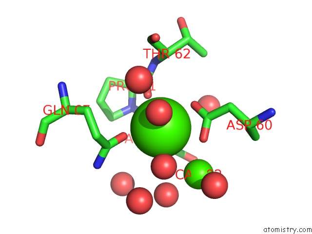
Calcium binding site 1 out of 4 in 4oy7
Go back to
Calcium binding site 1 out
of 4 in the Structure of Cellulose Active Lpmo CELS2 (SCLPMO10C) in Complex with Copper.
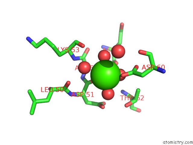
Mono view
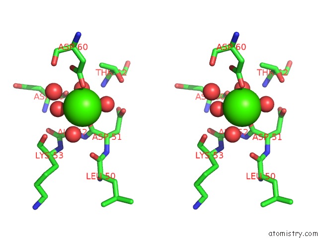
Stereo pair view
Mono view
Stereo pair view
A full contact list of Calcium with other atoms in the Ca binding
site number 1 of Structure of Cellulose Active Lpmo CELS2 (SCLPMO10C) in Complex with Copper. within 5.0Å range:
|
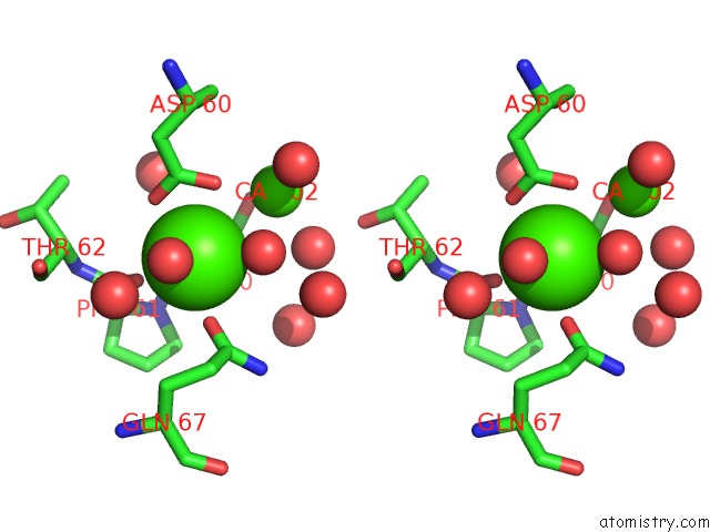
Calcium binding site 2 out of 4 in 4oy7
Go back to
Calcium binding site 2 out
of 4 in the Structure of Cellulose Active Lpmo CELS2 (SCLPMO10C) in Complex with Copper.
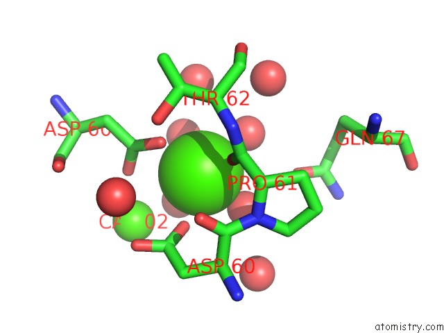
Mono view
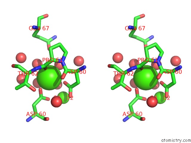
Stereo pair view
Mono view
Stereo pair view
A full contact list of Calcium with other atoms in the Ca binding
site number 2 of Structure of Cellulose Active Lpmo CELS2 (SCLPMO10C) in Complex with Copper. within 5.0Å range:
|
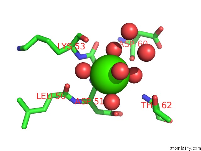
Calcium binding site 3 out of 4 in 4oy7
Go back to
Calcium binding site 3 out
of 4 in the Structure of Cellulose Active Lpmo CELS2 (SCLPMO10C) in Complex with Copper.
Mono view
Stereo pair view
Mono view
Stereo pair view
A full contact list of Calcium with other atoms in the Ca binding
site number 3 of Structure of Cellulose Active Lpmo CELS2 (SCLPMO10C) in Complex with Copper. within 5.0Å range:
|
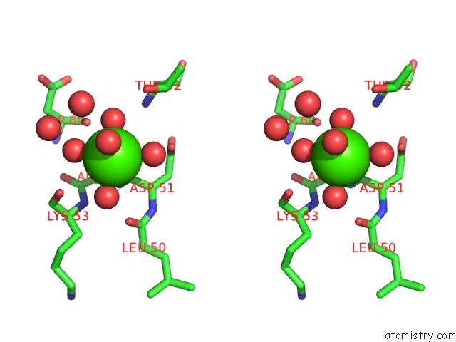
Calcium binding site 4 out of 4 in 4oy7
Go back to
Calcium binding site 4 out
of 4 in the Structure of Cellulose Active Lpmo CELS2 (SCLPMO10C) in Complex with Copper.
Mono view
Stereo pair view
Mono view
Stereo pair view
A full contact list of Calcium with other atoms in the Ca binding
site number 4 of Structure of Cellulose Active Lpmo CELS2 (SCLPMO10C) in Complex with Copper. within 5.0Å range:
|
Reference:
Z.Forsberg,
A.K.Mackenzie,
M.Srlie,
A.K.Rhr,
R.Helland,
A.S.Arvai,
G.Vaaje-Kolstad,
V.G.Eijsink.
Structural and Functional Characterization of A Conserved Pair of Bacterial Cellulose-Oxidizing Lytic Polysaccharide Monooxygenases. Proc.Natl.Acad.Sci.Usa V. 111 8446 2014.
ISSN: ESSN 1091-6490
PubMed: 24912171
DOI: 10.1073/PNAS.1402771111
Page generated: Wed Jul 9 01:11:17 2025
ISSN: ESSN 1091-6490
PubMed: 24912171
DOI: 10.1073/PNAS.1402771111
Last articles
Cl in 5JZNCl in 5JZB
Cl in 5JZL
Cl in 5JZ9
Cl in 5JZK
Cl in 5JY1
Cl in 5JYL
Cl in 5JXK
Cl in 5JXR
Cl in 5JXP