Calcium »
PDB 4opq-4p4f »
4p3q »
Calcium in PDB 4p3q: Room-Temperature Wt Dhfr, Time-Averaged Ensemble
Enzymatic activity of Room-Temperature Wt Dhfr, Time-Averaged Ensemble
All present enzymatic activity of Room-Temperature Wt Dhfr, Time-Averaged Ensemble:
1.5.1.3;
1.5.1.3;
Protein crystallography data
The structure of Room-Temperature Wt Dhfr, Time-Averaged Ensemble, PDB code: 4p3q
was solved by
D.A.Keedy,
H.Van Den Bedem,
J.S.Fraser,
with X-Ray Crystallography technique. A brief refinement statistics is given in the table below:
| Resolution Low / High (Å) | 41.34 / 1.35 |
| Space group | P 21 21 21 |
| Cell size a, b, c (Å), α, β, γ (°) | 34.320, 45.510, 98.910, 90.00, 90.00, 90.00 |
| R / Rfree (%) | 11.8 / 15.3 |
Calcium Binding Sites:
The binding sites of Calcium atom in the Room-Temperature Wt Dhfr, Time-Averaged Ensemble
(pdb code 4p3q). This binding sites where shown within
5.0 Angstroms radius around Calcium atom.
In total 2 binding sites of Calcium where determined in the Room-Temperature Wt Dhfr, Time-Averaged Ensemble, PDB code: 4p3q:
Jump to Calcium binding site number: 1; 2;
In total 2 binding sites of Calcium where determined in the Room-Temperature Wt Dhfr, Time-Averaged Ensemble, PDB code: 4p3q:
Jump to Calcium binding site number: 1; 2;
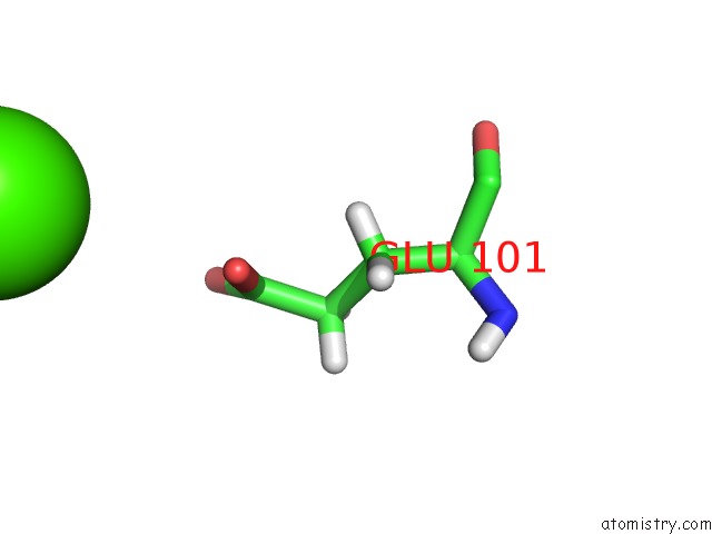
Calcium binding site 1 out of 2 in 4p3q
Go back to
Calcium binding site 1 out
of 2 in the Room-Temperature Wt Dhfr, Time-Averaged Ensemble
Mono view

Stereo pair view
Mono view
Stereo pair view
A full contact list of Calcium with other atoms in the Ca binding
site number 1 of Room-Temperature Wt Dhfr, Time-Averaged Ensemble within 5.0Å range:
|
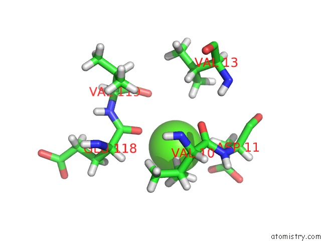
Calcium binding site 2 out of 2 in 4p3q
Go back to
Calcium binding site 2 out
of 2 in the Room-Temperature Wt Dhfr, Time-Averaged Ensemble
Mono view

Stereo pair view
Mono view
Stereo pair view
A full contact list of Calcium with other atoms in the Ca binding
site number 2 of Room-Temperature Wt Dhfr, Time-Averaged Ensemble within 5.0Å range:
|
Reference:
D.A.Keedy,
H.Van Den Bedem,
D.A.Sivak,
G.A.Petsko,
D.Ringe,
M.A.Wilson,
J.S.Fraser.
Crystal Cryocooling Distorts Conformational Heterogeneity in A Model Michaelis Complex of Dhfr. Structure V. 22 899 2014.
ISSN: ISSN 1878-4186
PubMed: 24882744
DOI: 10.1016/J.STR.2014.04.016
Page generated: Sun Jul 14 11:39:05 2024
ISSN: ISSN 1878-4186
PubMed: 24882744
DOI: 10.1016/J.STR.2014.04.016
Last articles
Zn in 9MJ5Zn in 9HNW
Zn in 9G0L
Zn in 9FNE
Zn in 9DZN
Zn in 9E0I
Zn in 9D32
Zn in 9DAK
Zn in 8ZXC
Zn in 8ZUF