Calcium »
PDB 4p4i-4pjs »
4p5f »
Calcium in PDB 4p5f: The Crystal Structure of Type III Effector Protein Xopq Complexed with Adenosine Diphosphate Ribose
Protein crystallography data
The structure of The Crystal Structure of Type III Effector Protein Xopq Complexed with Adenosine Diphosphate Ribose, PDB code: 4p5f
was solved by
S.Yu,
I.Hwang,
S.Rhee,
with X-Ray Crystallography technique. A brief refinement statistics is given in the table below:
| Resolution Low / High (Å) | 36.30 / 2.10 |
| Space group | P 21 21 21 |
| Cell size a, b, c (Å), α, β, γ (°) | 47.999, 73.808, 201.943, 90.00, 90.00, 90.00 |
| R / Rfree (%) | 20.2 / 25.3 |
Calcium Binding Sites:
The binding sites of Calcium atom in the The Crystal Structure of Type III Effector Protein Xopq Complexed with Adenosine Diphosphate Ribose
(pdb code 4p5f). This binding sites where shown within
5.0 Angstroms radius around Calcium atom.
In total 3 binding sites of Calcium where determined in the The Crystal Structure of Type III Effector Protein Xopq Complexed with Adenosine Diphosphate Ribose, PDB code: 4p5f:
Jump to Calcium binding site number: 1; 2; 3;
In total 3 binding sites of Calcium where determined in the The Crystal Structure of Type III Effector Protein Xopq Complexed with Adenosine Diphosphate Ribose, PDB code: 4p5f:
Jump to Calcium binding site number: 1; 2; 3;
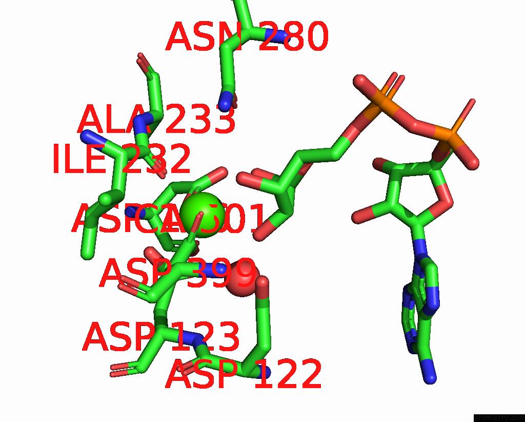
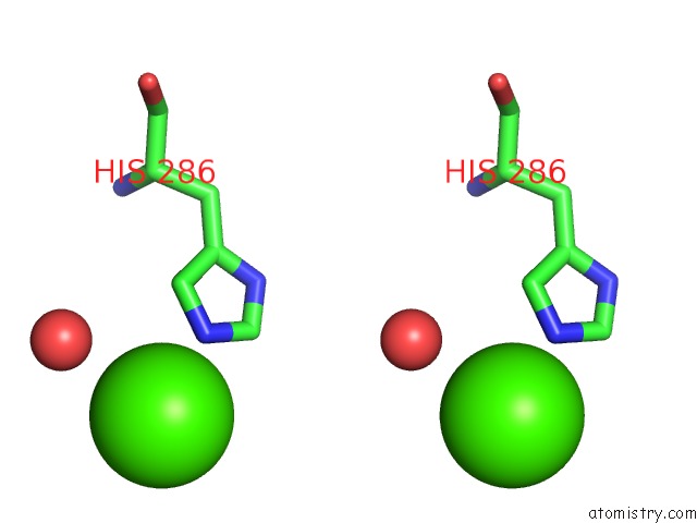
Calcium binding site 1 out of 3 in 4p5f
Go back to
Calcium binding site 1 out
of 3 in the The Crystal Structure of Type III Effector Protein Xopq Complexed with Adenosine Diphosphate Ribose

Mono view
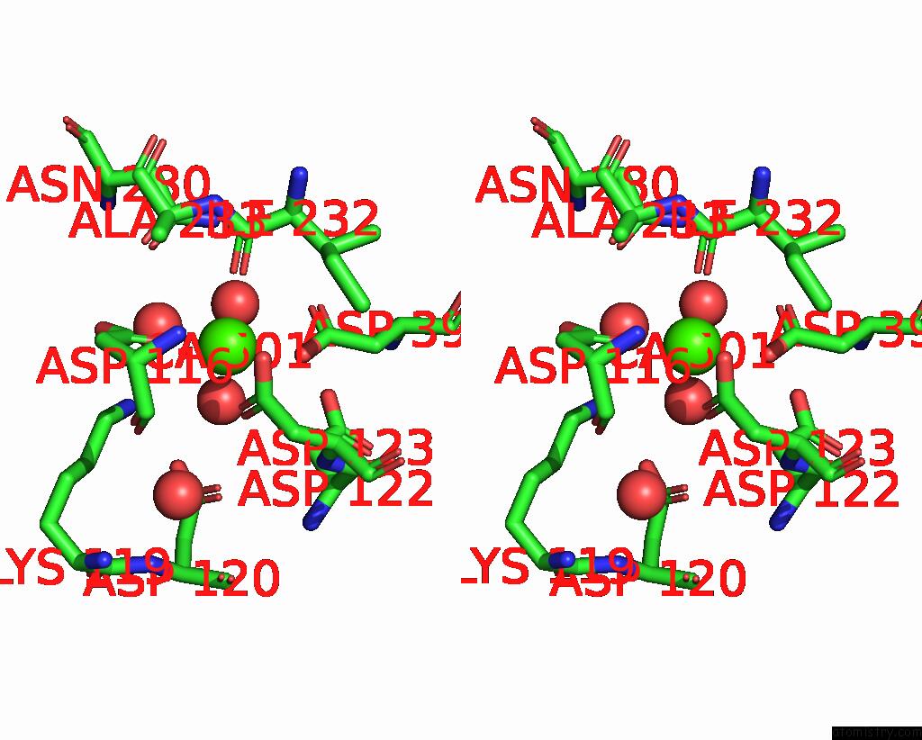
Stereo pair view
Mono view
Stereo pair view
A full contact list of Calcium with other atoms in the Ca binding
site number 1 of The Crystal Structure of Type III Effector Protein Xopq Complexed with Adenosine Diphosphate Ribose within 5.0Å range:
|
Calcium binding site 2 out of 3 in 4p5f
Go back to
Calcium binding site 2 out
of 3 in the The Crystal Structure of Type III Effector Protein Xopq Complexed with Adenosine Diphosphate Ribose
Mono view
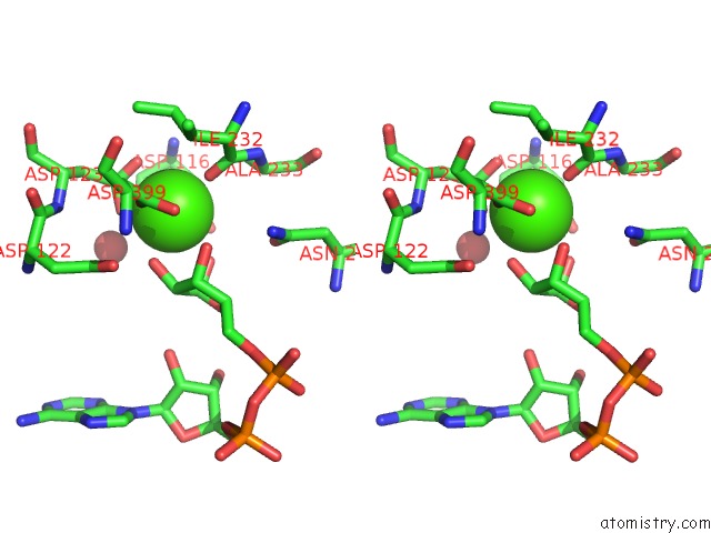
Stereo pair view
Mono view
Stereo pair view
A full contact list of Calcium with other atoms in the Ca binding
site number 2 of The Crystal Structure of Type III Effector Protein Xopq Complexed with Adenosine Diphosphate Ribose within 5.0Å range:
|
Calcium binding site 3 out of 3 in 4p5f
Go back to
Calcium binding site 3 out
of 3 in the The Crystal Structure of Type III Effector Protein Xopq Complexed with Adenosine Diphosphate Ribose
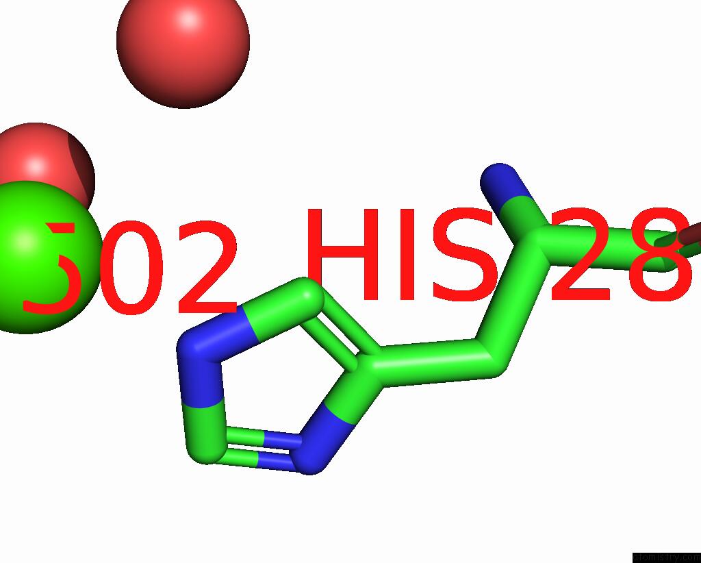
Mono view
Stereo pair view
Mono view
Stereo pair view
A full contact list of Calcium with other atoms in the Ca binding
site number 3 of The Crystal Structure of Type III Effector Protein Xopq Complexed with Adenosine Diphosphate Ribose within 5.0Å range:
|
Reference:
S.Yu,
I.Hwang,
S.Rhee.
The Crystal Structure of Type III Effector Protein Xopq From Xanthomonas Oryzae Complexed with Adenosine Diphosphate Ribose. Proteins V. 82 2910 2014.
ISSN: ESSN 1097-0134
PubMed: 25079351
DOI: 10.1002/PROT.24656
Page generated: Sun Jul 14 11:39:51 2024
ISSN: ESSN 1097-0134
PubMed: 25079351
DOI: 10.1002/PROT.24656
Last articles
Zn in 9JYWZn in 9IR4
Zn in 9IR3
Zn in 9GMX
Zn in 9GMW
Zn in 9JEJ
Zn in 9ERF
Zn in 9ERE
Zn in 9EGV
Zn in 9EGW