Calcium »
PDB 4p4i-4pjs »
4pe1 »
Calcium in PDB 4pe1: Crystal Structure of Calcium-Loaded S100B Bound to SC124
Protein crystallography data
The structure of Crystal Structure of Calcium-Loaded S100B Bound to SC124, PDB code: 4pe1
was solved by
M.C.Cavalier,
A.D.Pierce,
P.T.Wilder,
D.Neau,
E.A.Toth,
D.J.Weber,
with X-Ray Crystallography technique. A brief refinement statistics is given in the table below:
| Resolution Low / High (Å) | 35.35 / 1.58 |
| Space group | P 1 21 1 |
| Cell size a, b, c (Å), α, β, γ (°) | 34.649, 56.463, 48.269, 90.00, 110.10, 90.00 |
| R / Rfree (%) | 21.3 / 25.3 |
Calcium Binding Sites:
The binding sites of Calcium atom in the Crystal Structure of Calcium-Loaded S100B Bound to SC124
(pdb code 4pe1). This binding sites where shown within
5.0 Angstroms radius around Calcium atom.
In total 4 binding sites of Calcium where determined in the Crystal Structure of Calcium-Loaded S100B Bound to SC124, PDB code: 4pe1:
Jump to Calcium binding site number: 1; 2; 3; 4;
In total 4 binding sites of Calcium where determined in the Crystal Structure of Calcium-Loaded S100B Bound to SC124, PDB code: 4pe1:
Jump to Calcium binding site number: 1; 2; 3; 4;
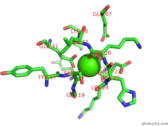
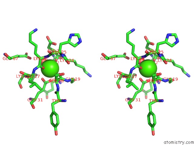
Calcium binding site 1 out of 4 in 4pe1
Go back to
Calcium binding site 1 out
of 4 in the Crystal Structure of Calcium-Loaded S100B Bound to SC124
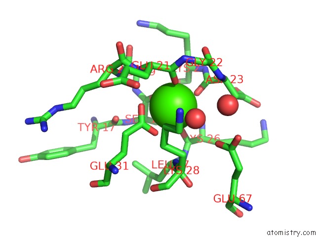
Mono view
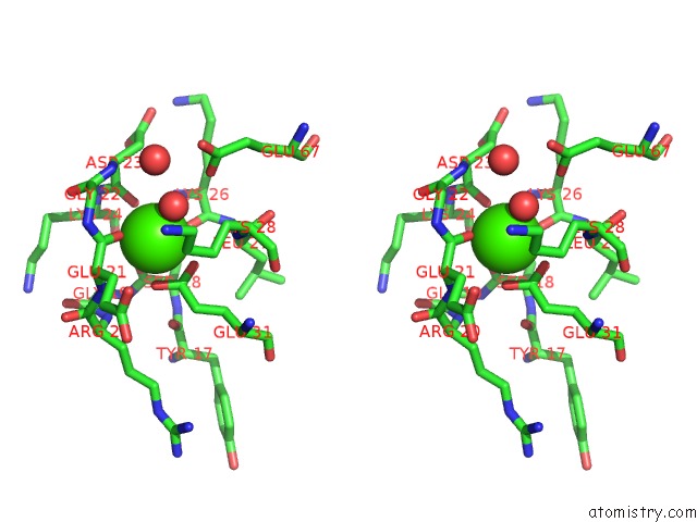
Stereo pair view
Mono view
Stereo pair view
A full contact list of Calcium with other atoms in the Ca binding
site number 1 of Crystal Structure of Calcium-Loaded S100B Bound to SC124 within 5.0Å range:
|
Calcium binding site 2 out of 4 in 4pe1
Go back to
Calcium binding site 2 out
of 4 in the Crystal Structure of Calcium-Loaded S100B Bound to SC124
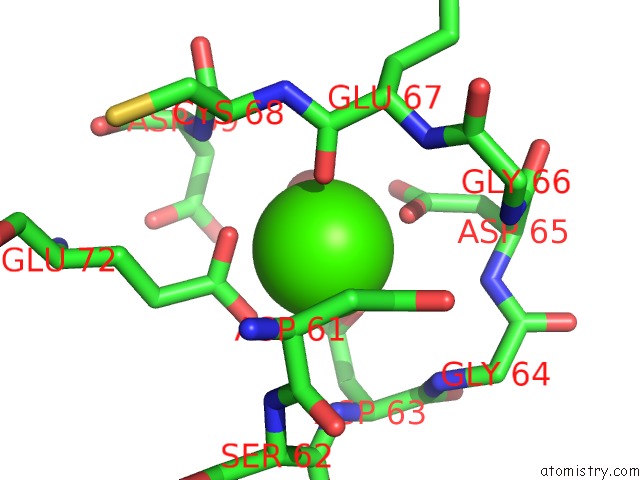
Mono view
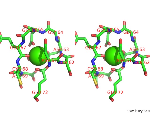
Stereo pair view
Mono view
Stereo pair view
A full contact list of Calcium with other atoms in the Ca binding
site number 2 of Crystal Structure of Calcium-Loaded S100B Bound to SC124 within 5.0Å range:
|
Calcium binding site 3 out of 4 in 4pe1
Go back to
Calcium binding site 3 out
of 4 in the Crystal Structure of Calcium-Loaded S100B Bound to SC124
Mono view
Stereo pair view
Mono view
Stereo pair view
A full contact list of Calcium with other atoms in the Ca binding
site number 3 of Crystal Structure of Calcium-Loaded S100B Bound to SC124 within 5.0Å range:
|
Calcium binding site 4 out of 4 in 4pe1
Go back to
Calcium binding site 4 out
of 4 in the Crystal Structure of Calcium-Loaded S100B Bound to SC124
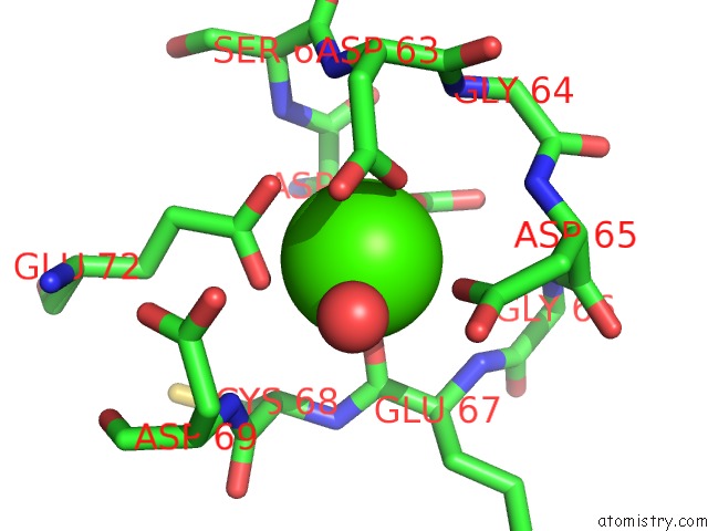
Mono view
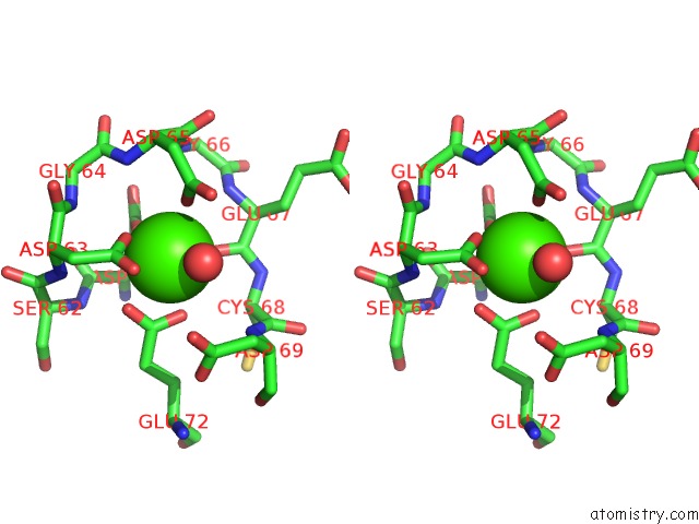
Stereo pair view
Mono view
Stereo pair view
A full contact list of Calcium with other atoms in the Ca binding
site number 4 of Crystal Structure of Calcium-Loaded S100B Bound to SC124 within 5.0Å range:
|
Reference:
M.C.Cavalier,
A.D.Pierce,
P.T.Wilder,
M.J.Alasady,
K.G.Hartman,
D.B.Neau,
T.L.Foley,
A.Jadhav,
D.J.Maloney,
A.Simeonov,
E.A.Toth,
D.J.Weber.
Covalent Small Molecule Inhibitors of Ca(2+)-Bound S100B. Biochemistry V. 53 6628 2014.
ISSN: ISSN 0006-2960
PubMed: 25268459
DOI: 10.1021/BI5005552
Page generated: Sun Jul 14 11:46:05 2024
ISSN: ISSN 0006-2960
PubMed: 25268459
DOI: 10.1021/BI5005552
Last articles
Zn in 9JYWZn in 9IR4
Zn in 9IR3
Zn in 9GMX
Zn in 9GMW
Zn in 9JEJ
Zn in 9ERF
Zn in 9ERE
Zn in 9EGV
Zn in 9EGW