Calcium »
PDB 4p4i-4pjs »
4pih »
Calcium in PDB 4pih: X-Ray Crystal Structure of the K33S Mutant of Ubiquitin
Protein crystallography data
The structure of X-Ray Crystal Structure of the K33S Mutant of Ubiquitin, PDB code: 4pih
was solved by
P.J.Loll,
P.J.Xu,
J.Schmidt,
S.L.Melideo,
with X-Ray Crystallography technique. A brief refinement statistics is given in the table below:
| Resolution Low / High (Å) | 23.25 / 1.50 |
| Space group | P 1 |
| Cell size a, b, c (Å), α, β, γ (°) | 27.350, 32.740, 40.340, 69.77, 72.55, 73.12 |
| R / Rfree (%) | 16.5 / 19 |
Other elements in 4pih:
The structure of X-Ray Crystal Structure of the K33S Mutant of Ubiquitin also contains other interesting chemical elements:
| Chlorine | (Cl) | 2 atoms |
Calcium Binding Sites:
The binding sites of Calcium atom in the X-Ray Crystal Structure of the K33S Mutant of Ubiquitin
(pdb code 4pih). This binding sites where shown within
5.0 Angstroms radius around Calcium atom.
In total 3 binding sites of Calcium where determined in the X-Ray Crystal Structure of the K33S Mutant of Ubiquitin, PDB code: 4pih:
Jump to Calcium binding site number: 1; 2; 3;
In total 3 binding sites of Calcium where determined in the X-Ray Crystal Structure of the K33S Mutant of Ubiquitin, PDB code: 4pih:
Jump to Calcium binding site number: 1; 2; 3;
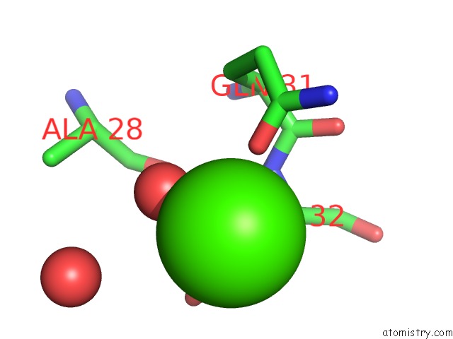
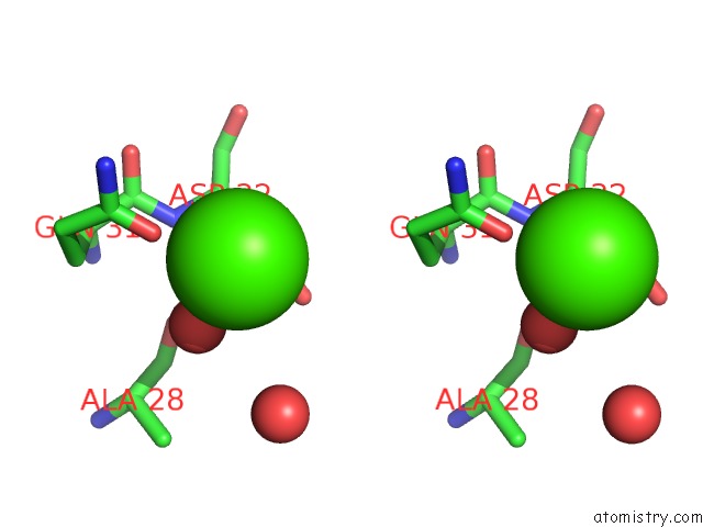
Calcium binding site 1 out of 3 in 4pih
Go back to
Calcium binding site 1 out
of 3 in the X-Ray Crystal Structure of the K33S Mutant of Ubiquitin
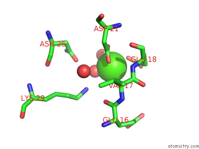
Mono view
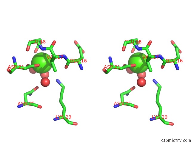
Stereo pair view
Mono view
Stereo pair view
A full contact list of Calcium with other atoms in the Ca binding
site number 1 of X-Ray Crystal Structure of the K33S Mutant of Ubiquitin within 5.0Å range:
|
Calcium binding site 2 out of 3 in 4pih
Go back to
Calcium binding site 2 out
of 3 in the X-Ray Crystal Structure of the K33S Mutant of Ubiquitin
Mono view
Stereo pair view
Mono view
Stereo pair view
A full contact list of Calcium with other atoms in the Ca binding
site number 2 of X-Ray Crystal Structure of the K33S Mutant of Ubiquitin within 5.0Å range:
|
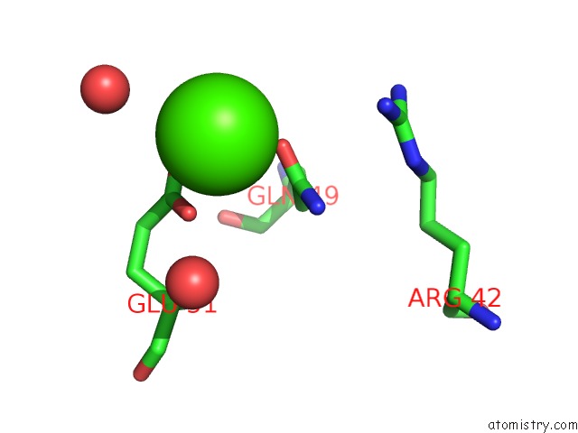
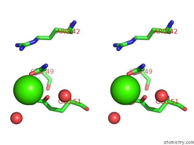
Calcium binding site 3 out of 3 in 4pih
Go back to
Calcium binding site 3 out
of 3 in the X-Ray Crystal Structure of the K33S Mutant of Ubiquitin
Mono view
Stereo pair view
Mono view
Stereo pair view
A full contact list of Calcium with other atoms in the Ca binding
site number 3 of X-Ray Crystal Structure of the K33S Mutant of Ubiquitin within 5.0Å range:
|
Reference:
P.J.Loll,
P.Xu,
J.T.Schmidt,
S.L.Melideo.
Enhancing Ubiquitin Crystallization Through Surface-Entropy Reduction. Acta Crystallogr.,Sect.F V. 70 1434 2014.
ISSN: ESSN 2053-230X
PubMed: 25286958
DOI: 10.1107/S2053230X14019244
Page generated: Sun Jul 14 11:54:24 2024
ISSN: ESSN 2053-230X
PubMed: 25286958
DOI: 10.1107/S2053230X14019244
Last articles
Zn in 9J0NZn in 9J0O
Zn in 9J0P
Zn in 9FJX
Zn in 9EKB
Zn in 9C0F
Zn in 9CAH
Zn in 9CH0
Zn in 9CH3
Zn in 9CH1