Calcium »
PDB 4pkg-4q1l »
4pwd »
Calcium in PDB 4pwd: Crystal Structure of Hiv-1 Reverse Transcriptase in Complex with Bulge-Rna/Dna and Nevirapine
Enzymatic activity of Crystal Structure of Hiv-1 Reverse Transcriptase in Complex with Bulge-Rna/Dna and Nevirapine
All present enzymatic activity of Crystal Structure of Hiv-1 Reverse Transcriptase in Complex with Bulge-Rna/Dna and Nevirapine:
2.7.7.49; 2.7.7.7; 3.1.13.2; 3.1.26.13;
2.7.7.49; 2.7.7.7; 3.1.13.2; 3.1.26.13;
Protein crystallography data
The structure of Crystal Structure of Hiv-1 Reverse Transcriptase in Complex with Bulge-Rna/Dna and Nevirapine, PDB code: 4pwd
was solved by
K.Das,
S.E.Martinez,
E.Arnold,
with X-Ray Crystallography technique. A brief refinement statistics is given in the table below:
| Resolution Low / High (Å) | 44.68 / 3.00 |
| Space group | P 1 21 1 |
| Cell size a, b, c (Å), α, β, γ (°) | 89.684, 130.765, 141.866, 90.00, 100.75, 90.00 |
| R / Rfree (%) | 22.1 / 29.1 |
Calcium Binding Sites:
The binding sites of Calcium atom in the Crystal Structure of Hiv-1 Reverse Transcriptase in Complex with Bulge-Rna/Dna and Nevirapine
(pdb code 4pwd). This binding sites where shown within
5.0 Angstroms radius around Calcium atom.
In total 2 binding sites of Calcium where determined in the Crystal Structure of Hiv-1 Reverse Transcriptase in Complex with Bulge-Rna/Dna and Nevirapine, PDB code: 4pwd:
Jump to Calcium binding site number: 1; 2;
In total 2 binding sites of Calcium where determined in the Crystal Structure of Hiv-1 Reverse Transcriptase in Complex with Bulge-Rna/Dna and Nevirapine, PDB code: 4pwd:
Jump to Calcium binding site number: 1; 2;
Calcium binding site 1 out of 2 in 4pwd
Go back to
Calcium binding site 1 out
of 2 in the Crystal Structure of Hiv-1 Reverse Transcriptase in Complex with Bulge-Rna/Dna and Nevirapine
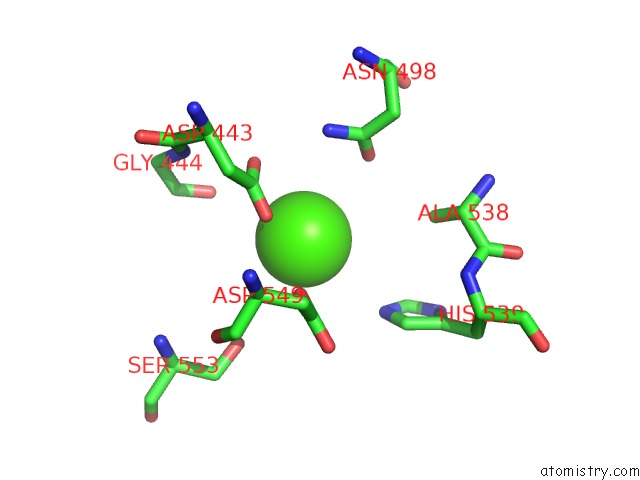
Mono view
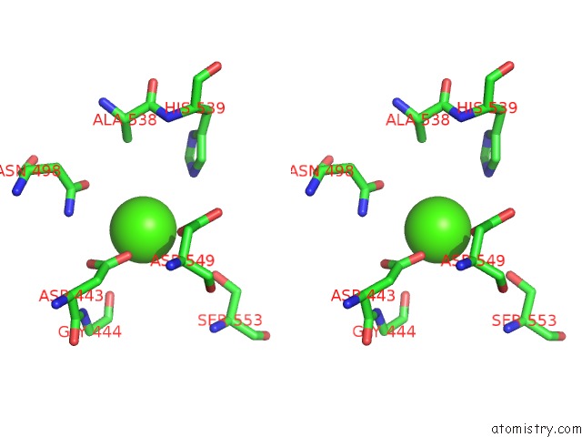
Stereo pair view
Mono view
Stereo pair view
A full contact list of Calcium with other atoms in the Ca binding
site number 1 of Crystal Structure of Hiv-1 Reverse Transcriptase in Complex with Bulge-Rna/Dna and Nevirapine within 5.0Å range:
|
Calcium binding site 2 out of 2 in 4pwd
Go back to
Calcium binding site 2 out
of 2 in the Crystal Structure of Hiv-1 Reverse Transcriptase in Complex with Bulge-Rna/Dna and Nevirapine
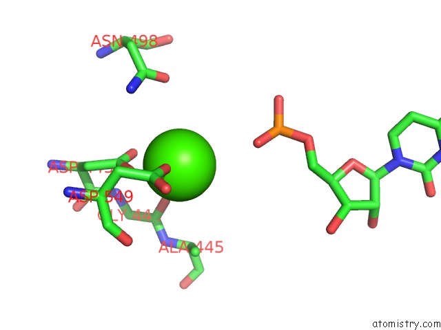
Mono view
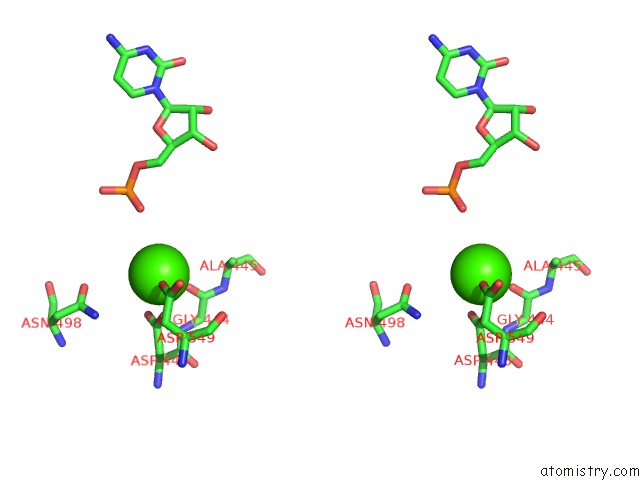
Stereo pair view
Mono view
Stereo pair view
A full contact list of Calcium with other atoms in the Ca binding
site number 2 of Crystal Structure of Hiv-1 Reverse Transcriptase in Complex with Bulge-Rna/Dna and Nevirapine within 5.0Å range:
|
Reference:
K.Das,
S.E.Martinez,
R.P.Bandwar,
E.Arnold.
Structures of Hiv-1 Rt-Rna/Dna Ternary Complexes with Datp and Nevirapine Reveal Conformational Flexibility of Rna/Dna: Insights Into Requirements For Rnase H Cleavage. Nucleic Acids Res. V. 42 8125 2014.
ISSN: ISSN 0305-1048
PubMed: 24880687
DOI: 10.1093/NAR/GKU487
Page generated: Sun Jul 14 12:15:08 2024
ISSN: ISSN 0305-1048
PubMed: 24880687
DOI: 10.1093/NAR/GKU487
Last articles
Zn in 9J0NZn in 9J0O
Zn in 9J0P
Zn in 9FJX
Zn in 9EKB
Zn in 9C0F
Zn in 9CAH
Zn in 9CH0
Zn in 9CH3
Zn in 9CH1