Calcium »
PDB 4pkg-4q1l »
4pwg »
Calcium in PDB 4pwg: Crystal Structure of V30M Mutant Human Transthyretin Complexed with Caffeic Acid Ethyl Ester
Protein crystallography data
The structure of Crystal Structure of V30M Mutant Human Transthyretin Complexed with Caffeic Acid Ethyl Ester, PDB code: 4pwg
was solved by
T.Yokoyama,
Y.Kosaka,
M.Mizuguchi,
with X-Ray Crystallography technique. A brief refinement statistics is given in the table below:
| Resolution Low / High (Å) | 30.39 / 1.80 |
| Space group | P 21 21 2 |
| Cell size a, b, c (Å), α, β, γ (°) | 43.190, 85.541, 63.771, 90.00, 90.00, 90.00 |
| R / Rfree (%) | 18.9 / 24.3 |
Calcium Binding Sites:
The binding sites of Calcium atom in the Crystal Structure of V30M Mutant Human Transthyretin Complexed with Caffeic Acid Ethyl Ester
(pdb code 4pwg). This binding sites where shown within
5.0 Angstroms radius around Calcium atom.
In total only one binding site of Calcium was determined in the Crystal Structure of V30M Mutant Human Transthyretin Complexed with Caffeic Acid Ethyl Ester, PDB code: 4pwg:
In total only one binding site of Calcium was determined in the Crystal Structure of V30M Mutant Human Transthyretin Complexed with Caffeic Acid Ethyl Ester, PDB code: 4pwg:
Calcium binding site 1 out of 1 in 4pwg
Go back to
Calcium binding site 1 out
of 1 in the Crystal Structure of V30M Mutant Human Transthyretin Complexed with Caffeic Acid Ethyl Ester
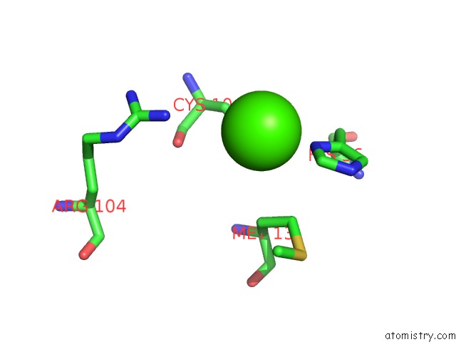
Mono view
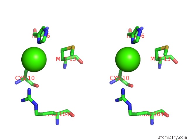
Stereo pair view
Mono view
Stereo pair view
A full contact list of Calcium with other atoms in the Ca binding
site number 1 of Crystal Structure of V30M Mutant Human Transthyretin Complexed with Caffeic Acid Ethyl Ester within 5.0Å range:
|
Reference:
T.Yokoyama,
Y.Kosaka,
M.Mizuguchi.
Inhibitory Activities of Propolis and Its Promising Component, Caffeic Acid Phenethyl Ester, Against Amyloidogenesis of Human Transthyretin J.Med.Chem. V. 57 8928 2014.
ISSN: ISSN 0022-2623
PubMed: 25314129
DOI: 10.1021/JM500997M
Page generated: Sun Jul 14 12:15:48 2024
ISSN: ISSN 0022-2623
PubMed: 25314129
DOI: 10.1021/JM500997M
Last articles
Zn in 9J0NZn in 9J0O
Zn in 9J0P
Zn in 9FJX
Zn in 9EKB
Zn in 9C0F
Zn in 9CAH
Zn in 9CH0
Zn in 9CH3
Zn in 9CH1