Calcium »
PDB 4pkg-4q1l »
4q1l »
Calcium in PDB 4q1l: Crystal Structure of Leucurolysin-A Complexed with An Endogenous Tripeptide (Qsw).
Protein crystallography data
The structure of Crystal Structure of Leucurolysin-A Complexed with An Endogenous Tripeptide (Qsw)., PDB code: 4q1l
was solved by
R.N.Ferreira,
E.O.F.Sanchez,
A.M.C.Pimenta,
R.A.P.Nagem,
with X-Ray Crystallography technique. A brief refinement statistics is given in the table below:
| Resolution Low / High (Å) | 32.10 / 1.90 |
| Space group | P 21 21 21 |
| Cell size a, b, c (Å), α, β, γ (°) | 44.350, 58.408, 76.842, 90.00, 90.00, 90.00 |
| R / Rfree (%) | 15.3 / 19.2 |
Other elements in 4q1l:
The structure of Crystal Structure of Leucurolysin-A Complexed with An Endogenous Tripeptide (Qsw). also contains other interesting chemical elements:
| Zinc | (Zn) | 1 atom |
Calcium Binding Sites:
The binding sites of Calcium atom in the Crystal Structure of Leucurolysin-A Complexed with An Endogenous Tripeptide (Qsw).
(pdb code 4q1l). This binding sites where shown within
5.0 Angstroms radius around Calcium atom.
In total only one binding site of Calcium was determined in the Crystal Structure of Leucurolysin-A Complexed with An Endogenous Tripeptide (Qsw)., PDB code: 4q1l:
In total only one binding site of Calcium was determined in the Crystal Structure of Leucurolysin-A Complexed with An Endogenous Tripeptide (Qsw)., PDB code: 4q1l:
Calcium binding site 1 out of 1 in 4q1l
Go back to
Calcium binding site 1 out
of 1 in the Crystal Structure of Leucurolysin-A Complexed with An Endogenous Tripeptide (Qsw).
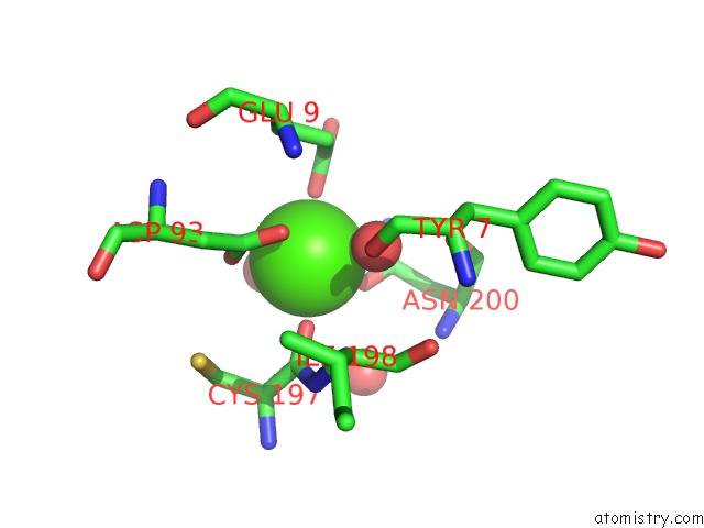
Mono view
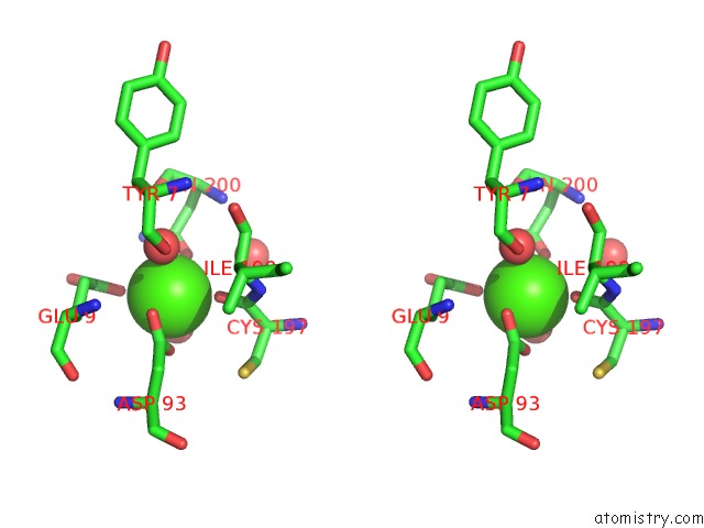
Stereo pair view
Mono view
Stereo pair view
A full contact list of Calcium with other atoms in the Ca binding
site number 1 of Crystal Structure of Leucurolysin-A Complexed with An Endogenous Tripeptide (Qsw). within 5.0Å range:
|
Reference:
R.N.Ferreira,
B.Rates,
M.Richardson,
B.G.Guimaraes,
E.O.F.Sanchez,
A.M.C.Pimenta,
R.A.P.Nagem.
Crystal Structure of Leucurolysin-A Complexed with An Endogenous Tripeptide (Qsw). To Be Published.
Page generated: Wed Jul 9 01:38:18 2025
Last articles
Cl in 8BQQCl in 8BQT
Cl in 8BRE
Cl in 8BQH
Cl in 8BQP
Cl in 8BOU
Cl in 8BOD
Cl in 8BOW
Cl in 8BOL
Cl in 8BO8