Calcium »
PDB 4q1o-4qn4 »
4q6m »
Calcium in PDB 4q6m: Structural Analysis of the Apo-Form of Helicobacter Pylori CSD4, A D, L-Carboxypeptidase
Protein crystallography data
The structure of Structural Analysis of the Apo-Form of Helicobacter Pylori CSD4, A D, L-Carboxypeptidase, PDB code: 4q6m
was solved by
H.S.Kim,
J.Kim,
H.N.Im,
D.R.An,
M.Lee,
D.Hesek,
S.Mobashery,
J.Y.Kim,
K.Cho,
H.J.Yoon,
B.W.Han,
B.I.Lee,
S.W.Suh,
with X-Ray Crystallography technique. A brief refinement statistics is given in the table below:
| Resolution Low / High (Å) | 29.81 / 1.60 |
| Space group | P 21 21 21 |
| Cell size a, b, c (Å), α, β, γ (°) | 53.141, 66.549, 144.053, 90.00, 90.00, 90.00 |
| R / Rfree (%) | 19.4 / 22 |
Calcium Binding Sites:
The binding sites of Calcium atom in the Structural Analysis of the Apo-Form of Helicobacter Pylori CSD4, A D, L-Carboxypeptidase
(pdb code 4q6m). This binding sites where shown within
5.0 Angstroms radius around Calcium atom.
In total 3 binding sites of Calcium where determined in the Structural Analysis of the Apo-Form of Helicobacter Pylori CSD4, A D, L-Carboxypeptidase, PDB code: 4q6m:
Jump to Calcium binding site number: 1; 2; 3;
In total 3 binding sites of Calcium where determined in the Structural Analysis of the Apo-Form of Helicobacter Pylori CSD4, A D, L-Carboxypeptidase, PDB code: 4q6m:
Jump to Calcium binding site number: 1; 2; 3;
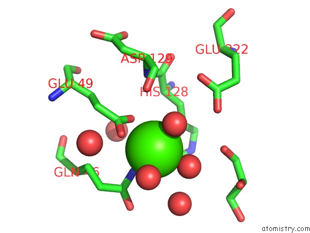
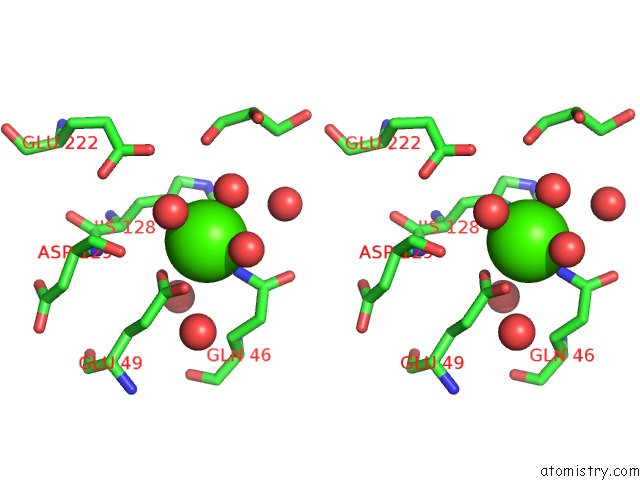
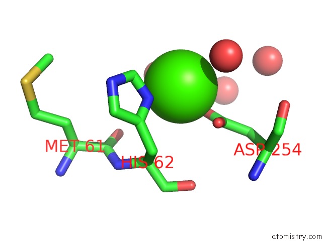
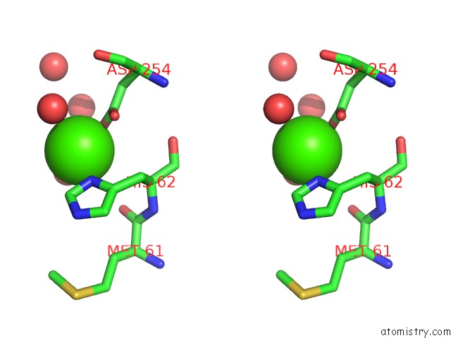
Calcium binding site 1 out of 3 in 4q6m
Go back to
Calcium binding site 1 out
of 3 in the Structural Analysis of the Apo-Form of Helicobacter Pylori CSD4, A D, L-Carboxypeptidase
Mono view
Stereo pair view
Mono view
Stereo pair view
A full contact list of Calcium with other atoms in the Ca binding
site number 1 of Structural Analysis of the Apo-Form of Helicobacter Pylori CSD4, A D, L-Carboxypeptidase within 5.0Å range:
|
Calcium binding site 2 out of 3 in 4q6m
Go back to
Calcium binding site 2 out
of 3 in the Structural Analysis of the Apo-Form of Helicobacter Pylori CSD4, A D, L-Carboxypeptidase
Mono view
Stereo pair view
Mono view
Stereo pair view
A full contact list of Calcium with other atoms in the Ca binding
site number 2 of Structural Analysis of the Apo-Form of Helicobacter Pylori CSD4, A D, L-Carboxypeptidase within 5.0Å range:
|
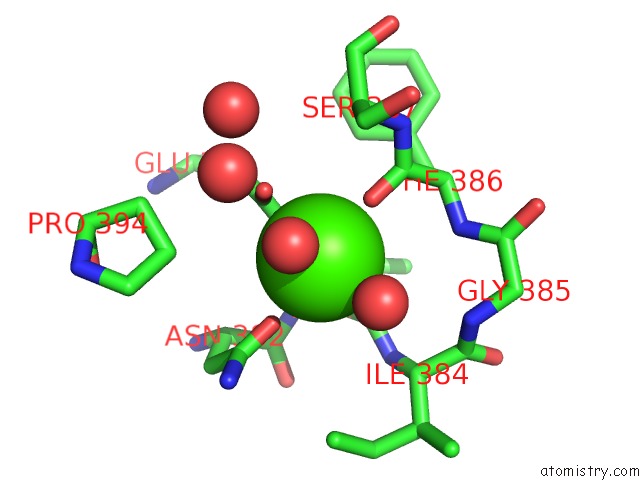
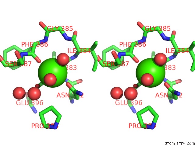
Calcium binding site 3 out of 3 in 4q6m
Go back to
Calcium binding site 3 out
of 3 in the Structural Analysis of the Apo-Form of Helicobacter Pylori CSD4, A D, L-Carboxypeptidase
Mono view
Stereo pair view
Mono view
Stereo pair view
A full contact list of Calcium with other atoms in the Ca binding
site number 3 of Structural Analysis of the Apo-Form of Helicobacter Pylori CSD4, A D, L-Carboxypeptidase within 5.0Å range:
|
Reference:
H.S.Kim,
J.Kim,
H.N.Im,
D.R.An,
M.Lee,
D.Hesek,
S.Mobashery,
J.Y.Kim,
K.Cho,
H.J.Yoon,
B.W.Han,
B.I.Lee,
S.W.Suh.
Structural Basis For the Recognition of Muramyltripeptide By Helicobacter Pylori CSD4, A D,L-Carboxypeptidase Controlling the Helical Cell Shape Acta Crystallogr.,Sect.D V. 70 2014.
ISSN: ESSN 1399-0047
DOI: 10.1107/S1399004714018732
Page generated: Sun Jul 14 12:21:30 2024
ISSN: ESSN 1399-0047
DOI: 10.1107/S1399004714018732
Last articles
Zn in 9JYWZn in 9IR4
Zn in 9IR3
Zn in 9GMX
Zn in 9GMW
Zn in 9JEJ
Zn in 9ERF
Zn in 9ERE
Zn in 9EGV
Zn in 9EGW