Calcium »
PDB 4q1o-4qn4 »
4qel »
Calcium in PDB 4qel: Crystal Structure of Benzoylformate Decarboxylase Mutant H70A
Enzymatic activity of Crystal Structure of Benzoylformate Decarboxylase Mutant H70A
All present enzymatic activity of Crystal Structure of Benzoylformate Decarboxylase Mutant H70A:
4.1.1.7;
4.1.1.7;
Protein crystallography data
The structure of Crystal Structure of Benzoylformate Decarboxylase Mutant H70A, PDB code: 4qel
was solved by
F.H.Andrews,
M.P.Rogers,
H.R.Brodkin,
M.J.Mcleish,
with X-Ray Crystallography technique. A brief refinement statistics is given in the table below:
| Resolution Low / High (Å) | 24.29 / 1.43 |
| Space group | I 2 2 2 |
| Cell size a, b, c (Å), α, β, γ (°) | 80.978, 95.442, 137.197, 90.00, 90.00, 90.00 |
| R / Rfree (%) | 13 / 15.8 |
Other elements in 4qel:
The structure of Crystal Structure of Benzoylformate Decarboxylase Mutant H70A also contains other interesting chemical elements:
| Magnesium | (Mg) | 1 atom |
| Chlorine | (Cl) | 1 atom |
Calcium Binding Sites:
The binding sites of Calcium atom in the Crystal Structure of Benzoylformate Decarboxylase Mutant H70A
(pdb code 4qel). This binding sites where shown within
5.0 Angstroms radius around Calcium atom.
In total 3 binding sites of Calcium where determined in the Crystal Structure of Benzoylformate Decarboxylase Mutant H70A, PDB code: 4qel:
Jump to Calcium binding site number: 1; 2; 3;
In total 3 binding sites of Calcium where determined in the Crystal Structure of Benzoylformate Decarboxylase Mutant H70A, PDB code: 4qel:
Jump to Calcium binding site number: 1; 2; 3;
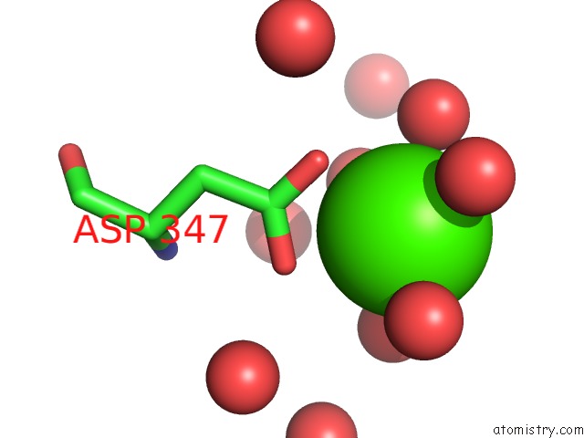
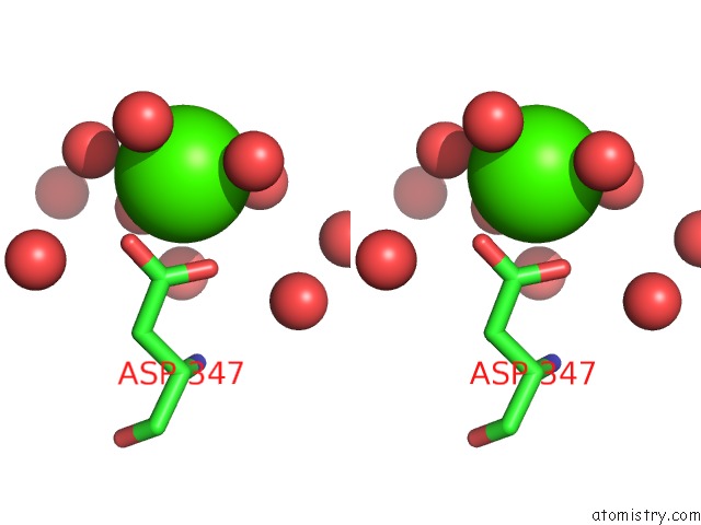
Calcium binding site 1 out of 3 in 4qel
Go back to
Calcium binding site 1 out
of 3 in the Crystal Structure of Benzoylformate Decarboxylase Mutant H70A
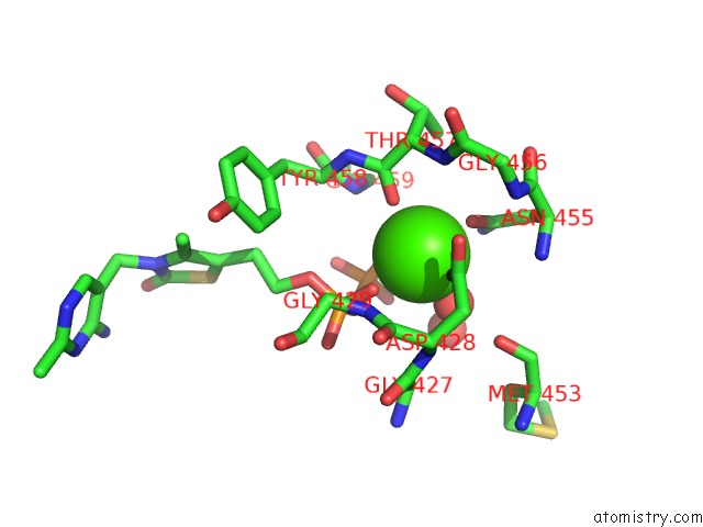
Mono view
Stereo pair view
Mono view
Stereo pair view
A full contact list of Calcium with other atoms in the Ca binding
site number 1 of Crystal Structure of Benzoylformate Decarboxylase Mutant H70A within 5.0Å range:
|
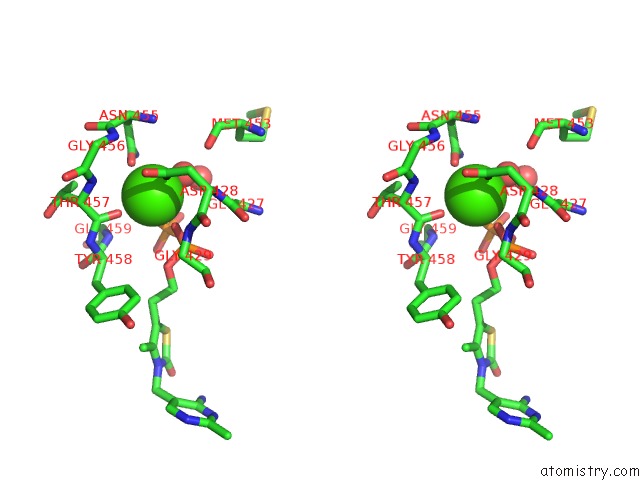
Calcium binding site 2 out of 3 in 4qel
Go back to
Calcium binding site 2 out
of 3 in the Crystal Structure of Benzoylformate Decarboxylase Mutant H70A
Mono view
Stereo pair view
Mono view
Stereo pair view
A full contact list of Calcium with other atoms in the Ca binding
site number 2 of Crystal Structure of Benzoylformate Decarboxylase Mutant H70A within 5.0Å range:
|
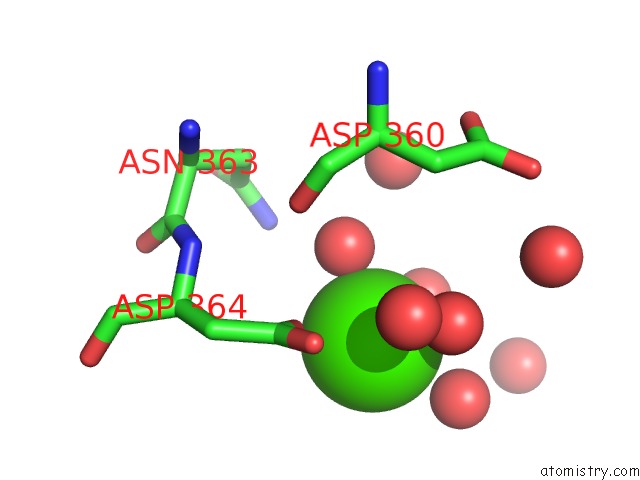
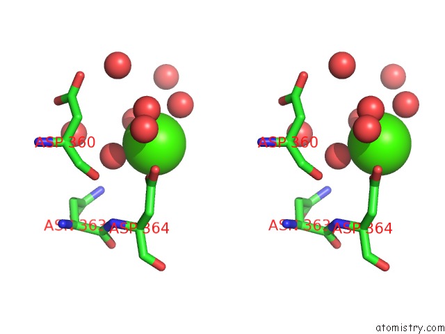
Calcium binding site 3 out of 3 in 4qel
Go back to
Calcium binding site 3 out
of 3 in the Crystal Structure of Benzoylformate Decarboxylase Mutant H70A
Mono view
Stereo pair view
Mono view
Stereo pair view
A full contact list of Calcium with other atoms in the Ca binding
site number 3 of Crystal Structure of Benzoylformate Decarboxylase Mutant H70A within 5.0Å range:
|
Reference:
F.H.Andrews,
M.P.Rogers,
H.R.Brodkin,
M.J.Mcleish.
Structural Investigation of Benzoylformate Decarboxylase Active Site Variants To Be Published.
Page generated: Sun Jul 14 12:24:44 2024
Last articles
Zn in 9JYWZn in 9IR4
Zn in 9IR3
Zn in 9GMX
Zn in 9GMW
Zn in 9JEJ
Zn in 9ERF
Zn in 9ERE
Zn in 9EGV
Zn in 9EGW