Calcium »
PDB 4q1o-4qn4 »
4qj4 »
Calcium in PDB 4qj4: Structure of A Fragment of Human Phospholipase C-BETA3 DELTA472-569, Bound to IP3 and in Complex with Galphaq
Enzymatic activity of Structure of A Fragment of Human Phospholipase C-BETA3 DELTA472-569, Bound to IP3 and in Complex with Galphaq
All present enzymatic activity of Structure of A Fragment of Human Phospholipase C-BETA3 DELTA472-569, Bound to IP3 and in Complex with Galphaq:
3.1.4.11;
3.1.4.11;
Protein crystallography data
The structure of Structure of A Fragment of Human Phospholipase C-BETA3 DELTA472-569, Bound to IP3 and in Complex with Galphaq, PDB code: 4qj4
was solved by
A.M.Lyon,
J.J.G.Tesmer,
with X-Ray Crystallography technique. A brief refinement statistics is given in the table below:
| Resolution Low / High (Å) | 29.40 / 3.30 |
| Space group | C 1 2 1 |
| Cell size a, b, c (Å), α, β, γ (°) | 201.923, 89.191, 92.639, 90.00, 101.70, 90.00 |
| R / Rfree (%) | 20.8 / 26.7 |
Other elements in 4qj4:
The structure of Structure of A Fragment of Human Phospholipase C-BETA3 DELTA472-569, Bound to IP3 and in Complex with Galphaq also contains other interesting chemical elements:
| Fluorine | (F) | 4 atoms |
| Magnesium | (Mg) | 1 atom |
| Aluminium | (Al) | 1 atom |
Calcium Binding Sites:
The binding sites of Calcium atom in the Structure of A Fragment of Human Phospholipase C-BETA3 DELTA472-569, Bound to IP3 and in Complex with Galphaq
(pdb code 4qj4). This binding sites where shown within
5.0 Angstroms radius around Calcium atom.
In total only one binding site of Calcium was determined in the Structure of A Fragment of Human Phospholipase C-BETA3 DELTA472-569, Bound to IP3 and in Complex with Galphaq, PDB code: 4qj4:
In total only one binding site of Calcium was determined in the Structure of A Fragment of Human Phospholipase C-BETA3 DELTA472-569, Bound to IP3 and in Complex with Galphaq, PDB code: 4qj4:
Calcium binding site 1 out of 1 in 4qj4
Go back to
Calcium binding site 1 out
of 1 in the Structure of A Fragment of Human Phospholipase C-BETA3 DELTA472-569, Bound to IP3 and in Complex with Galphaq
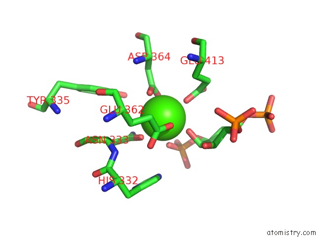
Mono view
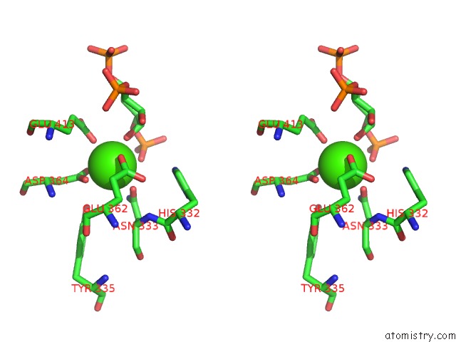
Stereo pair view
Mono view
Stereo pair view
A full contact list of Calcium with other atoms in the Ca binding
site number 1 of Structure of A Fragment of Human Phospholipase C-BETA3 DELTA472-569, Bound to IP3 and in Complex with Galphaq within 5.0Å range:
|
Reference:
A.M.Lyon,
J.A.Begley,
T.Manett,
J.J.G.Tesmer.
Molecular Mechanisms of Plcbeta Regulation. Structure 2014.
ISSN: ISSN 0969-2126
Page generated: Sun Jul 14 12:26:15 2024
ISSN: ISSN 0969-2126
Last articles
Zn in 9J0NZn in 9J0O
Zn in 9J0P
Zn in 9FJX
Zn in 9EKB
Zn in 9C0F
Zn in 9CAH
Zn in 9CH0
Zn in 9CH3
Zn in 9CH1